. 2021 Nov 12;16(11):e0259904.
doi: 10.1371/journal.pone.0259904. eCollection 2021.

Different virulence levels of Enterococcus cecorum strains in experimentally infected meat-type chickens

Affiliations

Different virulence levels of Enterococcus cecorum strains in experimentally infected meat-type chickens

Jana Schreier et al. PLoS One. .

Abstract

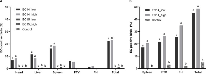
In recent years, pathogenic strains of Enterococcus cecorum (EC) have emerged as a causing agent of septicemia and skeletal infection in broiler chickens with a high economic impact worldwide. Although research has been conducted, many aspects of the pathogenesis of the EC-associated disease are still unknown. In the present study, an experimental infection model was established in broiler chickens. Two different EC strains (EC14 and EC15) were compared in two different concentrations of each strain (2 × 10⁶ and 2 × 10⁸ colony-forming units per milliliter (CFU/mL)) after oral infection of one-day-old chicks. Clinical signs and gross lesions of the EC-associated disease were monitored in the following seven weeks. Although both EC strains were originally isolated from clinical disease outbreaks and had a high embryonic lethality, only EC14 successfully induced the typical course of the EC-associated disease with characteristic clinical signs and gross lesions. In total, 23% of the birds in the two EC14-groups were EC-positive in extraintestinal organs on culture, and no differences were found between the two infectious doses. EC14 was frequently detected via real-time PCR in the free thoracic vertebra (FTV) and femoral heads without any detectable gross lesions. The number of EC positive spleens from infected broilers was comparable using bacterial isolation and a specific real-time PCR. Interestingly, EC15 was not detected in extraintestinal organs, although birds in the EC15 groups were colonized by EC in the ceca after experimental infection. The present study represents first proof that virulence differs among EC strains in experimentally infected chickens, and emphasizes the need to further characterize virulence factors and pathogenic mechanisms of EC. The strain EC14 at a dose of 106 CFU is suitable for reproduction of the EC-associated disease. The experimental infection model reported here provides the basis for further research on the EC pathogenesis and possible prevention and intervention strategies.


Conflict of interest statement

The authors have declared that no competing interests exist.

Figures

Fig 1
Fig 1
Clinical signs (A) and pathologic lesions (B) observed during the experiment. (A) Non-specific symptoms include depression, ruffled feathers, and closed eyes. Lameness includes all stages from mild to completely paralyzed. (B) Spondylitis FTV = abscess was found at the free thoracic vertebra, FH = femoral head. N = 147 per group in the EC-infected groups, N = 143 in the control group.
Fig 2
Fig 2. Clinical signs and pathology.
(A) Clinical signs of the septic phase of the EC-infection (study day 15, EC14_low). (B) Pericarditis due to EC (study day 25, EC14_low). (C) Clinical signs of the skeletal phase of the EC-infection. This broiler was sitting on hocks and unable to walk (study day 29, EC14_low). (D) Spondylitis at the FTV (arrow). The vertebral column was removed and split sagittally to expose the lesion (study day 30, EC14_high). (E) Clinical signs of the skeletal phase of the EC-infection. This broiler was lame due to femoral head osteomyelitis (study day 52, EC14_high). (F) Femoral head osteomyelitis (arrow). The femoral head was exarticulated by using a sterile scalpel and split sagittally (study day 43, EC14_low). EC was isolated in pure culture from the lesions in pictures B, D, and F.
Fig 3
Fig 3. Bacteriological examination via culture and real-time PCR.
(A) EC-positive birds on culture in %. (B) EC-positive birds in real-time PCR in %. Ct values below 36 were considered positive. Different letters indicate significant differences between the groups per organ (p ≤ 0.05). Comparison between the groups was made for each organ by using Fisher’s exact test. p-value adjustments for multiple testing were performed by using the Bonferroni-Holm correction method. Comparison between the groups was not done for culture results of the FTV and the femoral heads, because these samples were only taken when gross lesions were detected. N = 147 per group (EC-infected groups), N = 143 in the control group. FTV = free thoracic vertebra, FH = femoral heads.
Fig 4
Fig 4. Comparison of bacteriological examination of the spleen via culture and real-time PCR.
EC-positive spleens in groups EC14_low and EC14_high are shown per study day in %. Ct values below 36 were considered positive. All birds in the control group were found to be EC-negative in both detection methods. According to the Kappa coefficient, there was at least a substantial agreement (κ > 0.61) on the EC-status at all study days and McNemar’s test revealed no significant differences in distribution of results. N = 20 per group and study day.
Fig 5
Fig 5. Cecal colonization by EC.
Samples were analyzed via real-time PCR, and Ct values below 36 were considered positive. Different letters indicate significant differences between the groups per study day (p ≤ 0.05). Comparison between the groups was made for each study day by using Fisher’s exact test. p-value adjustments for multiple testing were performed by using the Bonferroni-Holm correction method. N = 20 per group and study day.

References

    1. Devriese LA, Dutta GN, Farrow JAE, Vandekerckhove A, Phillips BA. Streptococcus-Cecorum, a New Species Isolated from Chickens. Int J Syst Bacteriol. 1983;33(4):772–6. doi: 10.1099/00207713-33-4-772 WOS:A1983RL79800012. - DOI
    1. Devriese LA, Hommez J, Wijfels R, Haesebrouck F. Composition of the enterococcal and streptococcal intestinal flora of poultry. J Appl Bacteriol. 1991;71(1):46–50. Epub 1991/07/01. . - PubMed
    1. Martin LT, Martin MP, Barnes HJ. Experimental reproduction of enterococcal spondylitis in male broiler breeder chickens. Avian Dis. 2011;55(2):273–8. Epub 2011/07/29. doi: 10.1637/9614-121410-Reg.1 . - DOI - PubMed
    1. Devriese LA, Cauwerts K, Hermans K, Wood AM. Enterococcus cecorum septicemia as a cause of bone and joint lesions resulting in lameness in broiler chickens. Vlaams Diergeneeskundig Tijdschrift. 2002;71(3):219–21. WOS:000176537600005.
    1. Aitchison H, Poolman P, Coetzer M, Griffiths C, Jacobs J, Meyer M, et al.. Enterococcal-related vertebral osteoarthritis in South African broiler breeders: A case report. J S Afr Vet Assoc. 2014;85(1):1077. Epub 2015/02/17. doi: 10.4102/jsava.v85i1.1077 . - DOI - PubMed

MeSH terms