Review
. 2021 Dec:72:101510.
doi: 10.1016/j.arr.2021.101510. Epub 2021 Nov 9.

Iron homeostasis and organismal aging

Affiliations
Review

Iron homeostasis and organismal aging

Rola S Zeidan et al. Ageing Res Rev. 2021 Dec.

Abstract

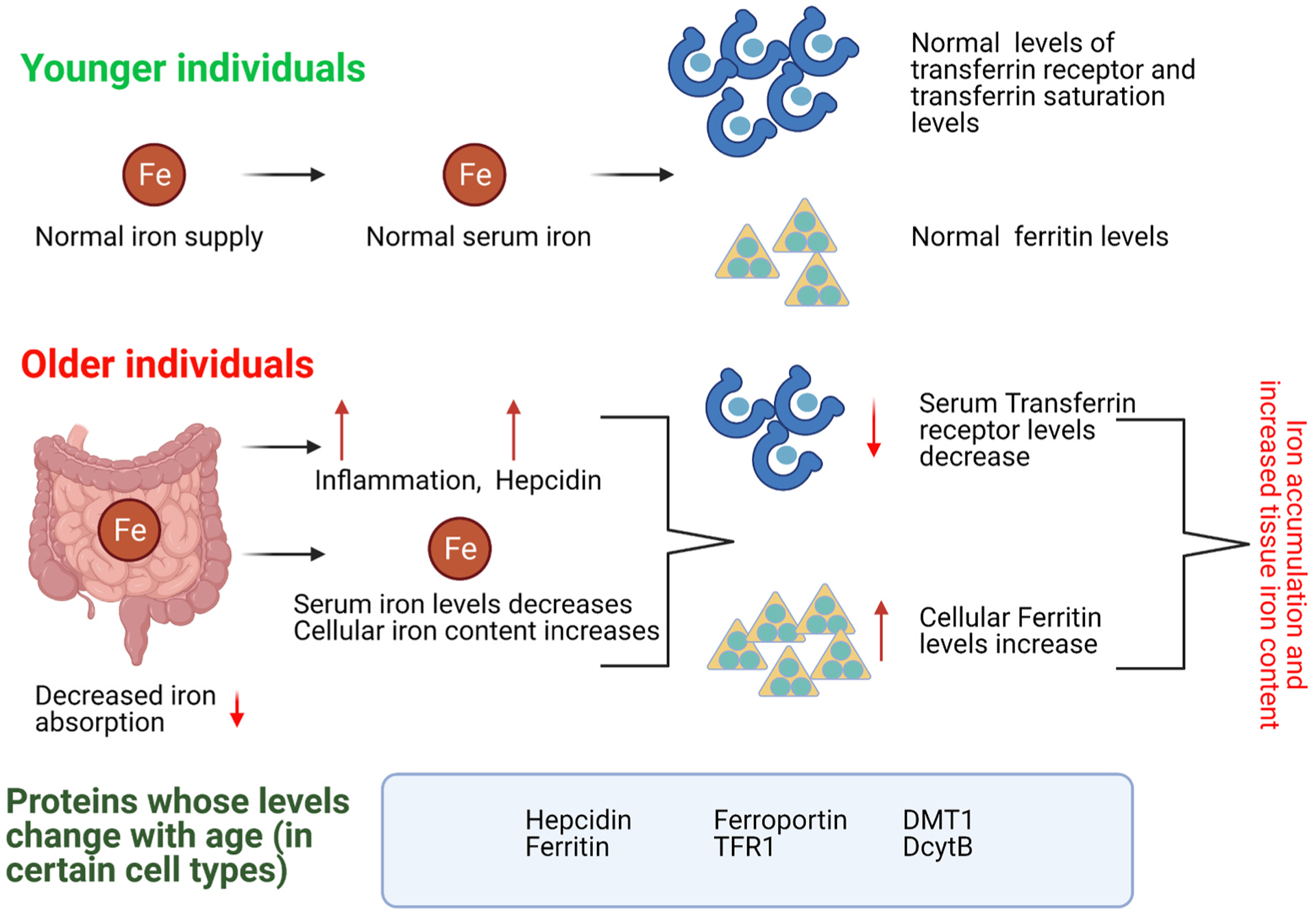
Iron is indispensable for normal body functions across species because of its critical roles in red blood cell function and many essential proteins and enzymes required for numerous physiological processes. Regulation of iron homeostasis is an intricate process involving multiple modulators at the systemic, cellular, and molecular levels. Interestingly, emerging evidence has demonstrated that many modulators of iron homeostasis contribute to organismal aging and longevity. On the other hand, the age-related dysregulation of iron homeostasis is often associated with multiple age-related pathologies including bone resorption and neurodegenerative diseases such as Alzheimer's disease. Thus, a thorough understanding on the interconnections between systemic and cellular iron balance and organismal aging may help decipher the etiologies of multiple age-related diseases, which could ultimately lead to developing therapeutic strategies to delay aging and treat various age-related diseases. Here we present the current understanding on the mechanisms of iron homeostasis. We also discuss the impacts of aging on iron homeostatic processes and how dysregulated iron metabolism may affect aging and organismal longevity.

Keywords: Aging; C. elegans; Drosophila; Homeostasis; Human diseases; Iron; Longevity.


Conflict of interest statement

Declaration of Competing Interest

The authors declare no competing interests.

Figures

Fig. 1.
Fig. 1.. Iron homeostasis is maintained by balancing iron uptake with turnover.
Aging and inflammation can disrupt iron homeostasis by enhancing iron accumulation in different tissues, which may lead to oxidative stress, neurodegeneration, organ damage, as well as cancer. Meanwhile, iron deficiencies are common with old age since dietary iron uptake via intestinal absorption becomes less efficient in the elderly.
Fig. 2.
Fig. 2.. The regulation of systemic iron homeostasis.
An increase in plasma (circulating) and tissue (stored) iron levels leads to production and secretion of the hormone hepcidin. Hepcidin decreases the levels of ferroportin (FPN) by direct binding, which leads to its internalization and degradation. The reduction of FPN causes a decrease in iron absorption at the intestinal level as well as a decrease in iron efflux by reticuloendothelial macrophages.
Fig. 3.
Fig. 3.. Important regulators involved in cellular iron homeostasis.
Non-heme iron can enter the cell in two forms: transferrin bound (TFB) and non-transferrin bound (aka NTBI). Ferrous iron or NTBI enters the cell through DMT1. Ferric iron is reduced to ferrous iron by DcytB, whereas ferrous iron is oxidized into ferric iron by the multicopper ferroxidases ceruloplasmin, hephaestin and HephL1. Ferric iron binds to transferrin that in turn binds to the transferrin receptor and gets internalized into the cell via endocytosis. Inside the internalized vesicles, iron is released and transformed into ferrous iron by STEAP3. Ferrous iron will then be exported out of the vesicle and into the cytoplasm by DMT1. Ferrous iron in the cytoplasm constitutes the labile iron pool (LIP), and it can be exported out of the cell by ferroportin, stored in the cell by ferritin, or utilized in different cellular processes. As for heme, it is synthesized in the mitochondria and can be exported to the outside of the cell by FLVCR1. Once in the cytoplasm, heme is cleaved by heme oxygenase (HO1) to release ferrous iron.
Fig. 4.
Fig. 4.. Age-related changes in iron homeostasis.
Transferrin receptor levels seem to decrease with age, while ferritin levels increase. Various studies with different animal models also suggest that depending on the organ studied, the levels of certain iron-regulating proteins can vary with age.
Fig. 5.
Fig. 5.. Iron metabolism and lifespan regulation in C. elegans.
A, excessive iron accumulation suppresses longevity in C. elegans, where ROS accumulation, ferroptosis, and cellular senescence may act as contributing factors. B, ISCU-1/ISCU, a central mitochondrial protein required for de novo biosynthesis of iron sulfur clusters, is essential for normal development during larval stages but accelerates aging during adulthood. C, suppressing the functions of ISCU-1 by iscu-1 RNAi extends lifespan and promotes stress resistance.
Fig. 6.
Fig. 6.. Organ-specific effects of age-related iron accumulation in humans.
Depending on the tissue and organ affected, age-related iron accumulation can lead to various pathologies in humans.

References

    1. Abbaspour N, Hurrell R, Kelishadi R, 2014. Review on iron and its importance for human health. J. Res. Med. Sci. J. Isfahan Univ. Med. Sci 19, 164–174. - PMC - PubMed
    1. Abdulzahra MS, Al-Hakeim HK, Ridha MM, 2011. Study of the effect of iron overload on the function of endocrine glands in male thalassemia patients. Asian J. Transfus. Sci 5, 127–131. 10.4103/0973-6247.83236. - DOI - PMC - PubMed
    1. Alleyne M, Horne MK, Miller JL, 2008. Individualized treatment for iron-deficiency anemia in adults. Am. J. Med 121, 943–948. 10.1016/j.amjmed.2008.07.012. - DOI - PMC - PubMed
    1. An P, Wu Q, Wang H, Guan Y, Mu M, Liao Y, Zhou D, Song P, Wang C, Meng L, Man Q, Li L, Zhang J, Wang F, 2012. TMPRSS6, but not TF, TFR2 or BMP2 variants are associated with increased risk of iron-deficiency anemia. Hum. Mol. Genet 21, 2124–2131. 10.1093/hmg/dds028. - DOI - PubMed
    1. Anderson CP, Shen M, Eisenstein RS, Leibold EA, 2012. Mammalian iron metabolism and its control by iron regulatory proteins. Biochim. Biophys. Acta BBA -Mol. Cell Res., Cell Biol. Met 1823, 1468–1483. 10.1016/j.bbamcr.2012.05.010. - DOI - PMC - PubMed

Publication types

LinkOut - more resources