. 2022 Jan;15(1):101265.
doi: 10.1016/j.tranon.2021.101265. Epub 2021 Nov 9.

Inhibitory monoclonal antibody targeting ADAM17 expressed on cancer cells

Affiliations

Inhibitory monoclonal antibody targeting ADAM17 expressed on cancer cells

Nayanendu Saha et al. Transl Oncol. 2022 Jan.

Abstract

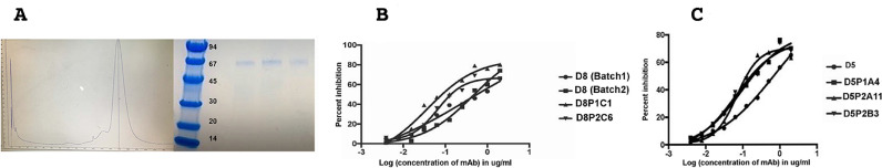
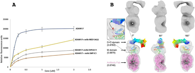
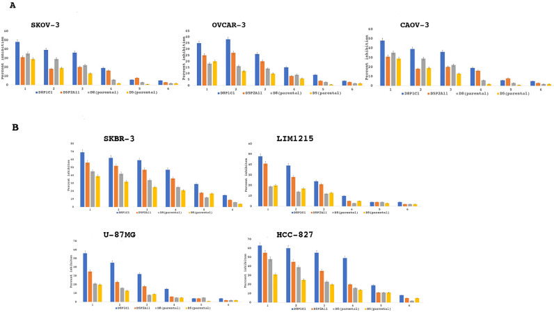
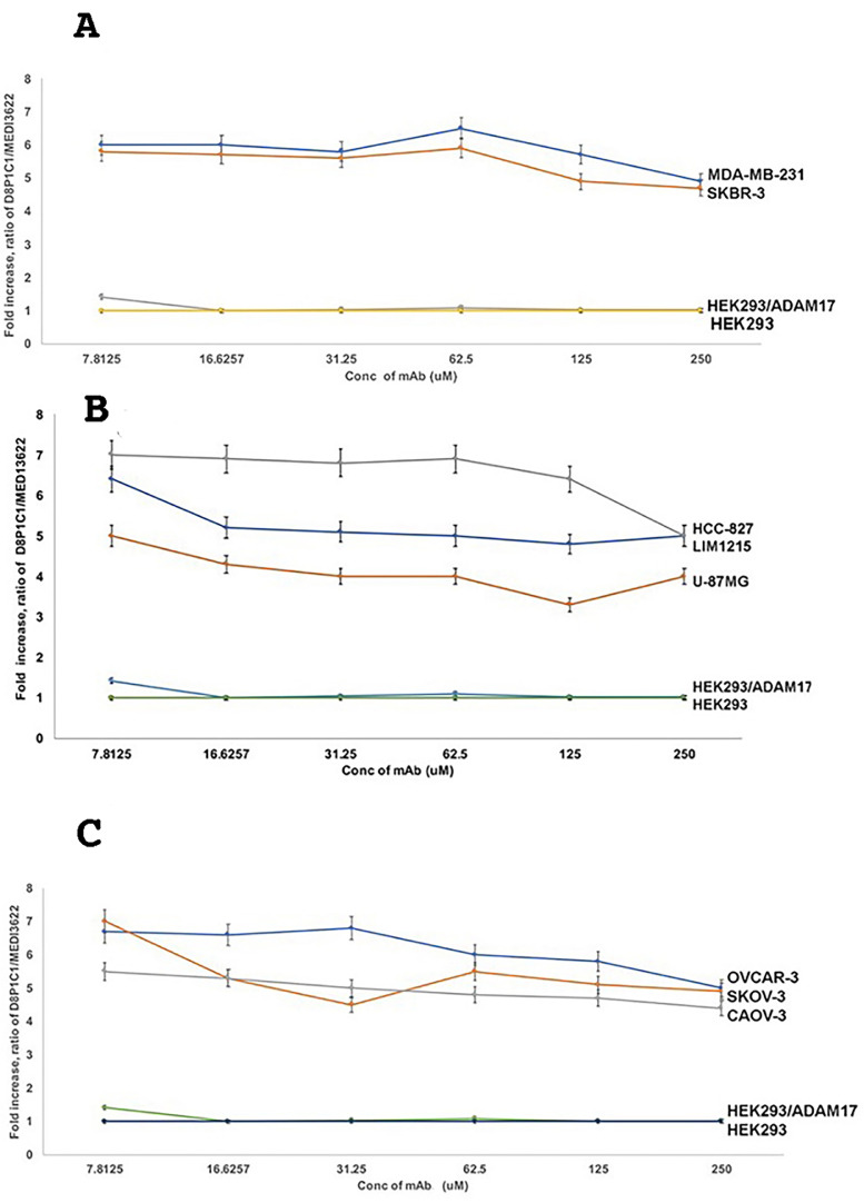
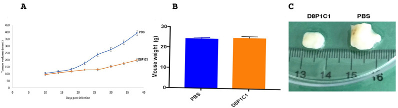
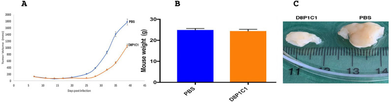
ADAM17 is upregulated in many cancers and in turn activates signaling pathways, including EGFR/ErbB, as well as those underlying resistance to targeted anti-EGFR therapies. Due to its central role in oncogenic pathways and drug resistance mechanisms, specific and efficacious monoclonal antibodies against ADAM17 could be useful for a broad patient population with solid tumors. Hence, we describe here an inhibitory anti-ADAM17 monoclonal antibody, named D8P1C1, that preferentially recognizes ADAM17 on cancer cells. D8P1C1 inhibits the catalytic activity of ADAM17 in a fluorescence-based peptide cleavage assay, as well as the proliferation of a range of cancer cell lines, including breast, ovarian, glioma, colon and the lung adenocarcinoma. In mouse models of triple-negative breast cancer and ovarian cancer, treatment with the mAb results in 78% and 45% tumor growth inhibition, respectively. Negative staining electron microscopy analysis of the ADAM17 ectodomain in complex with D8P1C1 reveals that the mAb binds the ADAM17 protease domain, consistent with its ability to inhibit the ADAM17 catalytic activity. Collectively, our results demonstrate the therapeutic potential of the D8P1C1 mAb to treat solid tumors.

Keywords: ADAM17; Cancer therapy; EGFR signaling; Monoclonal antibody.


Conflict of interest statement

The authors declare that they have no competing interest.

Figures

Fig. 1
Fig. 1
Antigen Expression and mAb Affinity Maturation. (A) SD-200 size-exclusion chromatography and SDS-PAGE profile of the purified ADAM17(ECD) construct. The molecular weights (kDa) of the protein standards are indicated. The three SDS-PAGE samples represent the three peak fractions from the SD-200 elute. The ADAM17(ECD) protein elutes as a monomer on the gel filtration chromatography column with a native molecular weight of 65 kDa. (B, C) Cell proliferation assays with MDA-MB-231 cells treated with five affinity matured clones of D5 and D8. The results represent mean of triplicate determinations and two independent experiments, and graphs show the effect of treatment of mAbs on MDA-MB-231 cells relative to the control (described in the Materials and Methods section, for IC50 and R-square values see Supplementary Table S1). D8 (Batch 1) was expressed and purified from Free Style™ 293 F cells and D8 (Batch 2) was expressed and purified from CHO cells.
Fig. 2
Fig. 2
The affinity matured D8P1C1 and D5P2A11 clones bind the ADAM17 metalloprotease domain and inhibit its proteolytic activity. (A) FRET-based peptide cleavage assays. The data represent mean of triplicate determinations and two independent experiments. Maximum dispersion was within 10% of the mean value. ADAM17-antibody complexes were formed at 1:1 molar ratio prior to the assay, and the assay was carried out in the presence of 50 μM of a fluorogenic peptide as described in the Materials and Methods section. Representative results of at least three independent experiments show effect of mAbs on substrate cleavage relative to the control (ADAM17 without the mAbs); mean ± SEM; P<0.001 by unpaired two-tailed Student's t-test, (ADAM17 without mAb vs ADAM17 with mAb). (B) Negative stain EM analysis and 3D reconstruction of the ADAM17 extracellular domain bound to a Fab derived from D8P1C1 shown in three perpendicular angles. The molecular volume rendering shows the electron density map (surface in gray) of the ADAM17/antibody complex (top), with crystal structures (bottom) of D + C domain (surface in rainbow color), M domain (cartoon in rainbow color) and antibody Fab (surface in magenta) [23, 24] fitted inside. Rainbow color follows the scheme of N-terminus in blue and C-terminus in red. Volume fitting correlation scores are shown in the parenthesis. A hypothetic linker between C-terminus of M domain and N-terminus of D + C domain is shown as dash line in the inset.
Fig. 3
Fig. 3
Alamar blue cell viability assays with multiple cancer cell lines. Percent growth inhibition is show for ovarian (SKOV-3, CAOV-3, OVCAR-3), breast (SKBR-3), colon (LIM1215), glioma (U87-MG) and non-small cell lung cancer (HCC-827) tumor cell lines treated with D8P1C1, D5P2A11, D8 or D5. The data represent mean of triplicate determinations and two independent experiments, and the bar plots show the effect of treatment of mAbs on cancer cells relative to the control, mean ± SEM (described in Materials and Methods).1: 20 µg/ml; 2: 10 µg/ml; 3: 5 µg/ml; 4: 2.5 µg/ml; 5: 1.25 µg/ml; 6: 0.625 µg/ml.
Fig. 4
Fig. 4
D8P1C1 preferentially binds to the activated, tumor-specific conformation of ADAM17. Cellular ELISA was performed to gage the binding of D8P1C1, relative to the binding of the control MED13622 mAb, to ADAM17 expressed on the cell surface of cancer cell lines: (A) breast, (B) colon, glioma and non-small cell lung, (C) ovarian, as well as HEK293 cells and HEK293 cells transfected with ADAM17. MED13622 binds equally well to the activated (tumor-associated) and the autoinhibited conformation of ADAM17. The graphs show the D8P1C1/MED13622 signal ratio relative to the D8P1C1/MED13622 signal ratio observed in the untransfected HEK293 cells. Specifically on the Y axes is plotted the value of A(D8P1C1)/A(MED13622)A(D8P1C1HEK)/A(MED13622HEK) Where A(D8P1C1-HEK) is the signal for D8P1C using the untransfected HEK293 cells; A(MED13622-HEK) is the signal for MED1362 using the untransfected HEK293 cells; A(D8P1C1-HEK) is the signal for D8P1C using the cells that are being evaluated; A(MED13622-HEK) is the signal for MED1362 using the cells being evaluated. D8P1C1 binds to ADAM17 on tumors approximately six-fold better than to ADAM17 on HEK2923 cells. The data represent triplicate determinations and two independent experiments, mean ± SEM; P < 0.001 by unpaired two-tailed Student's t-test (cancer cell lines vs HEK293 cells).
Fig. 5
Fig. 5
Anti-tumor effect of D8P1C1 in a triple-negative breast cancer xenograft model. 6–8 weeks old female athymic nude mice (n = 5) were used. 10 million MDA-MB-231 cells per mouse were introduced and each mouse was injected with D8P1C1 (i.p.) at a dose of 15 mg/kg, biweekly for 4 weeks. Sterile PBS was used as a control. Graphs show mean ± SEM, P< 0.001 by unpaired two-tailed Student's t-test, (mAb D8P1C1 vs PBS). D8P1C1 causes 78% tumor growth inhibition (A) with no loss in mouse weight (B) although it binds both human and mouse ADAM17. Panel (C) shows representative tumors excised from the treated and untreated animals.
Fig. 6
Fig. 6
Xenograft model using SKOV-3 ovarian cancer cells implanted into female NSG mice. The anti-ADAM17 mAb D8P1C1 causes 45 percent tumor growth inhibition at a dose of 60 mg/kg, administered bi-weekly for 4 weeks (A). 6–8 weeks old NSG mice (n = 5) were chosen. 10 million SKOV-3 cells per mouse were used and each mouse was injected with D8P1C1 (i.p.) at a dose of 60 mg/kg, biweekly for 4 weeks. Sterile PBS was used as a control. Graphs show mean ± SEM, P < 0.01 by unpaired two-tailed Student's t-test (mAb D8P1C1 vs PBS). Treatment with D8P1C1 did not affect mouse weight (B) although it binds both human and mouse ADAM17. Panel (C) shows representative tumors excised from the treated and untreated animals.

References

    1. Murphy G. The ADAMS: signalling scissors in the tumour microenvironment. Nat. Rev. Cancer. 2008;8:929–941. - PubMed
    1. Dreymueller D., Uhlig S., Ludwig A. ADAM-family metalloproteinases in lung inflammation: potential therapeutic targets. Am. J. Physiol.-Lung Cell. Mol. Physiol. 2015;308(4):L325–L343. - PubMed
    1. Pruessmeyer J., Ludwig A. The good, the bad and the ugly substrates for ADAM10 and ADAM17 in brain pathology, inflammation and cancer. Semin. Cell Dev. Biol. 2009;20(2):164–174. - PubMed
    1. Seals D.F., Courtneidge SA S.A. The ADAMs family of metalloproteases: multidomain proteins with multiple functions. Genes Dev. 2003;17(1):7–30. - PubMed
    1. Janes P.W., Saha N., Barton W.A., Kolev M.V., Wimmer-Kleikamp S.H., Nievergall E., Blobel C.P., Himanen J.P., Lackmann M., Nikolov D.B. Adam meets Eph: an ADAM substrate recognition module acts as a molecular switch for ephrin cleavage in trans. Cell. 2005;123(2):291–304. - PubMed