Review
. 2021 Oct 21;10(21):4841.
doi: 10.3390/jcm10214841.

Non-Adhesive Liquid Embolic Agents in Extra-Cranial District: State of the Art and Review of the Literature

Affiliations
Review

Non-Adhesive Liquid Embolic Agents in Extra-Cranial District: State of the Art and Review of the Literature

Filippo Piacentino et al. J Clin Med. .

Abstract

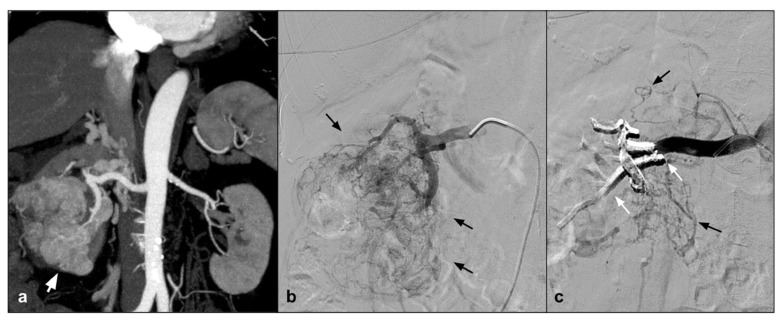
This review focuses on the use of "new" generation of non-adhesive liquid embolic agents (NALEA). In literature, non-adhesive liquid embolic agents have mainly been used in the cerebral district; however, multiple papers describing the use of NALEA in the extracranial district have been published recently and the aim of this review is to explore and analyze this field of application. There are a few NALEA liquids such as Onyx, Squid, and Phil currently available in the market, and they are used in the following applications: mainly arteriovenous malformations, endoleaks, visceral aneurysm or pseudoaneurysm, presurgical and hypervascular lesions embolization, and a niche of percutaneous approaches. These types of embolizing fluids can be used alone or in combination with other embolizing agents (such as coils or particles) so as to enhance its embolizing effect or improve its possible defects. The primary purpose of this paper is to evaluate the use of NALEAs, predominantly used alone, in elective embolization procedures. We did not attempt a meta-analysis due to the data heterogeneity, high number of case reports, and the lack of a consistent follow-up time period.

Keywords: EVOH; Onyx; Squid; artery embolization; non-adhesive liquid embolic agent.


Conflict of interest statement

The authors declare no conflict of interest.

Figures

Figure 1
Figure 1
(a). Coronal CEMRA MIP demonstrates the presence of voluminous AVM originating from the right uterine artery with aneurysmal venous outflow (white arrows); (b) DSA performed with a vertebral shape catheter in the ipsilateral hypogastric artery confirms the presence of the AVM highlighting the “nidus” (black arrows); (c) single-shot post-treatment fluoroscopy shows the inferior uterine artery treated (white arrow) with coils (not directly tributary of the AVM) and the presence of a cast of Squid 12 at the level of the middle and inferior uterine artery which were the arterial feeding vessels of the AVM (black arrows); (d) post-procedure DSA demonstrates the complete exclusion of the treated AVM (asterisk).
Figure 2
Figure 2
(a) Coronal CTA MIP demonstrates hypertrophic bronchial artery with tortuous course originating at 9:00 from the convexity of the aortic arch (white arrows); (b) DSA performed with Simmons 1 catheter at the level of the ostium of the bronchial artery confirms the presence of the hypertrophic vessel (black arrow) with a small tributary branch of the right upper lobe (arrow head); (c) single-shot fluoroscopy after embolization shows the presence of the microcatheter at the level of the proximal portion of the bronchial artery, with a Squid 12 cast completely occupying the main trunk (white arrows); (d) post-procedure DSA control demonstrates complete exclusion of the treated vessel (asterisk) with patency of the small branch for the right pulmonary upper lobe (white arrow).
Figure 3
Figure 3
(a) Axial CTA shows, the presence of Type II endoleak after EVAR, supplied by lumbar arteries (white arrow). (b) DSA performed with microcatheter positioned in a lumbar branch through the ilio-lumbar artery highlights the presence of hypertrophic lumbar circles (white arrow head) with sac refuelling (white arrow). (c) Post-procedure DSA shows the cast of Squid 12, which completely occupies the space of the endoleak in the sac (white arrow).
Figure 4
Figure 4
(a) Coronal CTA MIP demonstrates post surgical pseudoaneurysm of left gastric artery (white arrows). (b) DSA performed with Simmons 1 catheter at the level of the ostium of the celiac trunk confirms the presence of the pseudoaneurysm (white arrow). (c) DSA performed with microcatheter in the left gastric artery highlights a saccular dilation (white arrow) of the left gastric artery with regular patency of the efferent vessel (black arrow). (d) Post-procedure DSA control demonstrates complete exclusion of the treated PSA (asterisk) with an Onyx 18 cast completely occupying the malacic vessel performing an “endovascular ligature technique” (white arrow).
Figure 5
Figure 5
(a) Coronal CTA MIP highlights a large angiomyolipoma of the right kidney lower third (white arrow). (b) DSA performed with a direct injection in the right renal artery confirms the lesion with an abnormal arterial vascularization (black arrows). (c) A post procedure DSA check demonstrates the presence of the Squid 34 cast which completely occludes the tributary arteries of the lesion (white arrows) and preservation of the healthy renal parenchyma (black arrows).

References

    1. Kim P.H., Tsauo J., Shin J.H., Yun S.-C. Transcatheter Arterial Embolization of Gastrointestinal Bleeding with N-Butyl Cyanoacrylate: A Systematic Review and Meta-Analysis of Safety and Efficacy. J. Vasc. Interv. Radiol. 2017;28:522–531. doi: 10.1016/j.jvir.2016.12.1220. - DOI - PubMed
    1. Lucatelli P., Corona M., Teodoli L., Nardis P., Cannavale A., Rocco B., Trobiani C., Cipollari S., Zilahi de Gyurgyokai S., Bezzi M., et al. Use of Phil Embolic Agent for Bleeding in Non-Neurological Interventions. J. Clin. Med. 2021;10:701. doi: 10.3390/jcm10040701. - DOI - PMC - PubMed
    1. Alturki A.Y., Enriquez-Marulanda A., Schmalz P., Ogilvy C.S., Thomas A.J. Transarterial Onyx Embolization of Bilateral Transverse-Sigmoid Dural Arteriovenous Malformation with Transvenous Balloon Assist-Initial U.S. Experience with Copernic RC Venous Remodeling Balloon. World Neurosurg. 2018;109:398–402. doi: 10.1016/j.wneu.2017.10.083. - DOI - PubMed
    1. Nerva J.D., Barber J., Levitt M.R., Rockhill J.K., Hallam D.K., Ghodke B.V., Sekhar L.N., Kim L.J. Onyx Embolization Prior to Stereotactic Radiosurgery for Brain Arteriovenous Malformations: A Single-Center Treatment Algorithm. J. Neurointerv. Surg. 2018;10:258–267. doi: 10.1136/neurintsurg-2017-013084. - DOI - PubMed
    1. Singfer U., Hemelsoet D., Vanlangenhove P., Martens F., Verbeke L., Van Roost D., Defreyne L. Unruptured Brain Arteriovenous Malformations: Primary ONYX Embolization in ARUBA (A Randomized Trial of Unruptured Brain Arteriovenous Malformations)-Eligible Patients. Stroke. 2017;48:3393–3396. doi: 10.1161/STROKEAHA.117.018605. - DOI - PubMed

LinkOut - more resources