

Anti-Oxidation and Anti-Inflammatory Potency Evaluation of Ferulic Acid Derivatives Obtained through Virtual Screening
- PMID: 34768735
- PMCID: PMC8583578
- DOI: 10.3390/ijms222111305
Anti-Oxidation and Anti-Inflammatory Potency Evaluation of Ferulic Acid Derivatives Obtained through Virtual Screening
Abstract
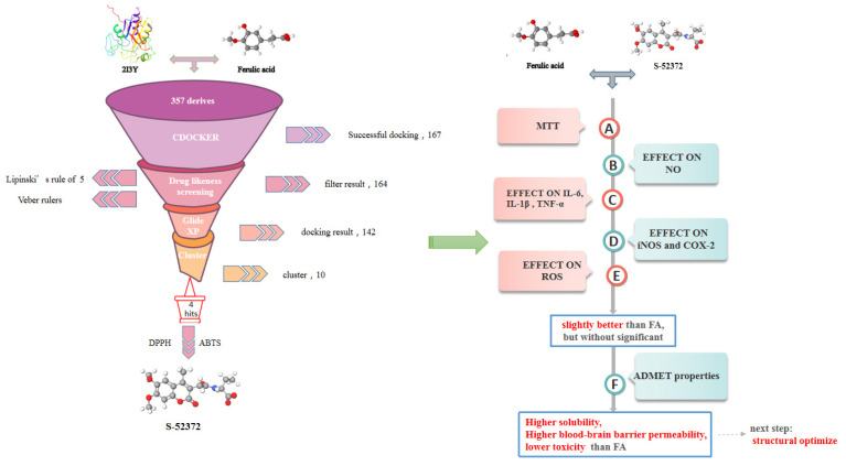
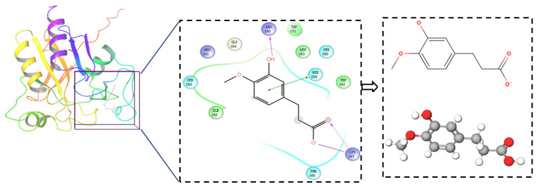
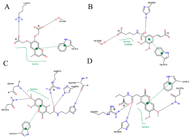
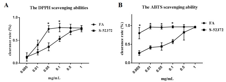
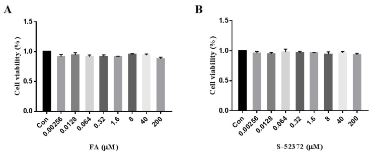
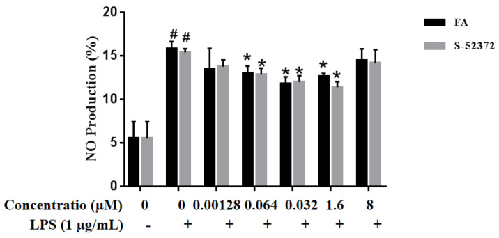
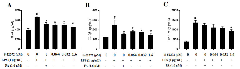
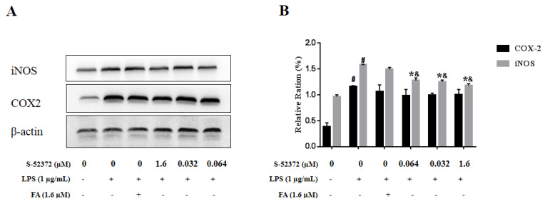
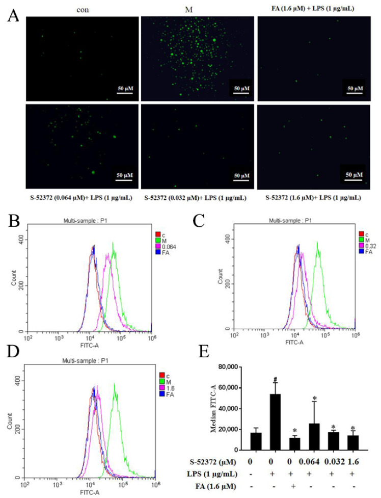
Various factors such as ultraviolet rays can cause a continuous threat to our skin, resulting in inflammation or oxidation problems. Ferulic acid (FA), with certain antioxidant and anti-inflammatory properties, is widely used in many cosmetics, even used to treat various diseases in the clinic. In this study, the FA structural skeleton was used to search for FA derivatives. Then, molecular docking, the rule of five, and Veber rules were performed to virtually screen compounds that can bind to proteins with a good drug likeness. DPPH and ABTS were used to evaluate their antioxidant potency and an MTT assay was employed to investigate the toxicities of the compounds, while Griess Reaction System and ELISA were used to judge the concentration variations of NO and different inflammatory factors (TNF-α, IL-1β, and IL-6). Western blotting featured nitric oxide synthase (iNOS) and cyclooxygenase-2 (COX-2) protein expression levels. The trend of the intracellular changes of reactive oxygen species (ROS) was detected by the DCFH-DA method and fluorescence staining. As a result, we found that the ferulic acid derivative S-52372 not only had certain scavenging effects on free radicals in biochemical experiments, but also prevented inflammation and oxidative stress in LPS-stimulated RAW264.7 cells in the cellular environment; intracellular ROS and inflammatory mediators, including iNOS, COX-2, TNF-α, and IL-6, were also suppressed. In a computer prediction, S-52372 owned better water solubility and lower toxicity than FA. This compound deserves further research to find an ideal FA derivative.
Keywords: anti-inflammation; antioxidant; ferulic acid; free radical scavenging; virtual screening.
Conflict of interest statement
The authors declare no conflict of interest.
Figures
Similar articles
-
Ferulic acid but not alpha-lipoic acid effectively protects THP-1-derived macrophages from oxidant and pro-inflammatory response to LPS.Immunopharmacol Immunotoxicol. 2017 Dec;39(6):330-337. doi: 10.1080/08923973.2017.1369100. Epub 2017 Sep 5. Immunopharmacol Immunotoxicol. 2017. PMID: 28872362
-
Quercetin and Quercitrin Attenuates the Inflammatory Response and Oxidative Stress in LPS-Induced RAW264.7 Cells: In Vitro Assessment and a Theoretical Model.Biomed Res Int. 2019 Oct 28;2019:7039802. doi: 10.1155/2019/7039802. eCollection 2019. Biomed Res Int. 2019. PMID: 31781635 Free PMC article.
-
DXXK exerts anti-inflammatory effects by inhibiting the lipopolysaccharide-induced NF-κB/COX-2 signalling pathway and the expression of inflammatory mediators.J Ethnopharmacol. 2016 Feb 3;178:199-208. doi: 10.1016/j.jep.2015.11.016. Epub 2015 Nov 10. J Ethnopharmacol. 2016. PMID: 26571085
-
Therapeutic Promises of Ferulic Acid and its Derivatives on Hepatic damage Related with Oxidative Stress and Inflammation: A Review with Mechanisms.Chem Biodivers. 2024 Jul;21(7):e202400443. doi: 10.1002/cbdv.202400443. Epub 2024 Jun 26. Chem Biodivers. 2024. PMID: 38757848 Review.
-
Comparison of in vitro tests for antioxidant and immunomodulatory capacities of compounds.Phytomedicine. 2014 Jan 15;21(2):164-71. doi: 10.1016/j.phymed.2013.08.008. Epub 2013 Sep 14. Phytomedicine. 2014. PMID: 24041614 Review.
Cited by
-
Antioxidant, Phytochemical, and Pharmacological Properties of Algerian Mentha aquatica Extracts.Antioxidants (Basel). 2024 Dec 11;13(12):1512. doi: 10.3390/antiox13121512. Antioxidants (Basel). 2024. PMID: 39765840 Free PMC article.
-
Unlocking the Bioactive Potential and Exploring Novel Applications for Portuguese Endemic Santolina impressa.Plants (Basel). 2024 Jul 15;13(14):1943. doi: 10.3390/plants13141943. Plants (Basel). 2024. PMID: 39065470 Free PMC article.
-
Impact of Protein Content on the Antioxidants, Anti-Inflammatory Properties and Glycemic Index of Wheat and Wheat Bran.Foods. 2022 Jul 11;11(14):2049. doi: 10.3390/foods11142049. Foods. 2022. PMID: 35885294 Free PMC article.
-
Structural Optimization of Cannabidiol as Multifunctional Cosmetic Raw Materials.Antioxidants (Basel). 2023 Jan 29;12(2):314. doi: 10.3390/antiox12020314. Antioxidants (Basel). 2023. PMID: 36829873 Free PMC article.
-
Molecular mechanism of ferulic acid and its derivatives in tumor progression.Pharmacol Rep. 2023 Aug;75(4):891-906. doi: 10.1007/s43440-023-00494-0. Epub 2023 May 18. Pharmacol Rep. 2023. PMID: 37202657 Free PMC article. Review.
References
-
- Ghezzi P., Floridi L., Boraschi D., Cuadrado A., Manda G., Levic S., D’Acquisto F., Hamilton A., Athersuch T.J., Selley L. Oxidative Stress and Inflammation Induced by Environmental and Psychological Stressors: A Biomarker Perspective. Antioxid. Redox Signal. 2018;28:852–872. doi: 10.1089/ars.2017.7147. - DOI - PubMed
-
- Akashi S., Saitoh S., Wakabayashi Y., Kikuchi T., Takamura N., Nagai Y., Kusumoto Y., Fukase K., Kusumoto S., Adachi Y., et al. Lipopolysaccharide interaction with cell surface Toll-like receptor 4-MD-2: Higher affinity than that with MD-2 or CD14. J. Exp. Med. 2003;198:1035–1042. doi: 10.1084/jem.20031076. - DOI - PMC - PubMed
MeSH terms
Substances
LinkOut - more resources
Full Text Sources
Research Materials
