. 2021 Oct 20;22(21):11328.
doi: 10.3390/ijms222111328.

Gastric Helicobacter suis Infection Partially Protects against Neurotoxicity in A 6-OHDA Parkinson's Disease Mouse Model

Affiliations

Gastric Helicobacter suis Infection Partially Protects against Neurotoxicity in A 6-OHDA Parkinson's Disease Mouse Model

Helena Berlamont et al. Int J Mol Sci. .

Abstract

The exact etiology of Parkinson's disease (PD) remains largely unknown, but more and more research suggests the involvement of the gut microbiota. Interestingly, idiopathic PD patients were shown to have at least a 10 times higher prevalence of Helicobacter suis (H. suis) DNA in gastric biopsies compared to control patients. H. suis is a zoonotic Helicobacter species that naturally colonizes the stomach of pigs and non-human primates but can be transmitted to humans. Here, we investigated the influence of a gastric H. suis infection on PD disease progression through a 6-hydroxydopamine (6-OHDA) mouse model. Therefore, mice with either a short- or long-term H. suis infection were stereotactically injected with 6-OHDA in the left striatum and sampled one week later. Remarkably, a reduced loss of dopaminergic neurons was seen in the H. suis/6-OHDA groups compared to the control/6-OHDA groups. Correspondingly, motor function of the H. suis-infected 6-OHDA mice was superior to that in the non-infected 6-OHDA mice. Interestingly, we also observed higher expression levels of antioxidant genes in brain tissue from H. suis-infected 6-OHDA mice, as a potential explanation for the reduced 6-OHDA-induced cell loss. Our data support an unexpected neuroprotective effect of gastric H. suis on PD pathology, mediated through changes in oxidative stress.

Keywords: 6-OHDA; Helicobacter; Parkinson’s disease; gut–brain axis; oxidative stress.


Conflict of interest statement

The authors declare no conflict of interest.

Figures

Figure 1
Figure 1
Experimental set-up to investigate the effect of Helicobacter suis (H. suis) infection in a 6-hydroxydopamine (6-OHDA) mouse model of Parkinson’s disease (PD). Animal experiments were carried out consisting of a short- and long-term infection group, respectively. C57BL/6OlaHsd mice were intragastrically inoculated at the age of 42 days with 300 µL of a stock solution containing 1 × 10⁸ viable H. suis HS1 bacteria/mL (n = 10 for short-term and n = 11 for long-term) or with the growth medium of the bacteria (i.e., Brucella broth (pH 5)) as negative controls (n = 10 for short-term and n = 9 for long-term). Twenty-one days post-infection (short-term group) and 500 days post-infection (long-term group), mice were injected in the left striatum with either 1 µL of 6-OHDA hydrochloride solution (16 µg/µL 6-OHDA in 0.2% ascorbic acid) or the vehicle solution (0.2% ascorbic acid in phosphate buffered saline). Seven days after intrastriatal injection, mice were sampled. Behavior and motor function tests were performed at two different timepoints. Baseline testing took place in the week before intrastriatal injection. The second testing took place 5–6 days after intrastriatal injection and 1–2 days prior to sampling.
Figure 2
Figure 2
Both short- and long-term gastric Helicobacter suis (H. suis) infection is associated with gastric colonization and inflammation. (A,B) H. suis colonization in the stomach (corpus and antrum) of mice infected with H. suis or the control broth for a short- (n = 9–11) (A) and long-term period (n = 10) (B), 7 days after instrastriatal injection with either 6-OHDA (black) or vehicle (white). (C,D) Gastric inflammation score according to the Updated Sydney System based on haematoxylin and eosin (H&E) staining of the stomach of control and H. suis-infected mice for short- (n = 9–11) (C) and long-term infection (n = 10) (D). (EH) Relative mRNA gene expression of the cytokine interleukin 1β (Il1β) and the chemokine keratinocyte chemoattractant (Kc) in the corpus of the stomach after short- (n = 9–11) (E) and long-term infection (n= 7–10) (F). Data were analyzed by the Mann–Whitney test. ** 0.001 ≤ p < 0.01; *** 0.0001 ≤ p < 0.001; **** p < 0.0001.
Figure 3
Figure 3
Gastric Helicobacter suis (H. suis) infection is associated with a limited decrease in integrity of the gastrointestinal barrier. (A,B) Relative mRNA gene expression of the mucin Muc13 in the corpus of the stomach of mice infected with H. suis or the control broth for a short- (n = 8–10) (A) or long-term period (n = 7–10) (B), 7 days after intrastriatal injection with either 6-OHDA (black) or vehicle (white). (CH) Relative expression of the tight junction genes claudin 5 (Cldn5) (C,D), zonula occludens 1 (Zo1) (E,F), and zonula occludens 3 (Zo3) (G,H) in the corpus of the stomach of mice infected with H. suis or the control broth for a short- (n = 9–11) (C,E,G) or long-term period (n = 8–10) (D,F,H). (I,J) Relative gastrointestinal permeability based on the 4 kDa FITC-dextran leakage assay of control mice compared to mice infected with H. suis for a short- (n = 8–9) (I) or long-term period (n = 9–10) (J). Data were analyzed by the Mann–Whitney test. * 0.01 ≤ p < 0.05; ** 0.001 ≤ p < 0.01; *** 0.0001 ≤ p < 0.001.
Figure 4
Figure 4
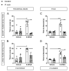
Helicobacter suis (H. suis)-infected mice are partially protected against the motor deficits induced by intrastriatal 6-hydroxydopamine (6-OHDA) injection. (A,B) Time (in seconds (s)) to cross the traversal beam, 7 days after intrastriatal injection with either 6-OHDA (n = 4–5) or vehicle (n = 5–6), of mice infected with H. suis (black) or the control broth (white) for a short-term (A) or long-term period (B). (C,D) Time (in seconds (s)) to descend the pole, 7 days after intrastriatal injection with either 6-OHDA (n = 4–5) or vehicle (n = 5–6), of mice infected with H. suis (black) or the control broth (white) for a short- (C) or long-term period (D). (E,F) Stride length of the right hind limb according to the footprint analysis, 7 days after intrastriatal injection with either 6-OHDA (n = 4–5) or vehicle (n = 5–6), of mice infected with H. suis (black) or the control broth (white) for a short-term period (E) or a long-term period (F). (G,H) Total amount of wall touches in the cylinder test, 7 days after intrastriatal injection with either 6-OHDA (n = 4–5) or vehicle (n = 5–6), of mice infected with H. suis (black) or the control broth (white) for a short- (A) or long-term period (B). * 0.01 ≤ p < 0.05; ** 0.001 ≤ p < 0.01; *** 0.0001 ≤ p < 0.001; **** p < 0.0001.
Figure 4
Figure 4
Helicobacter suis (H. suis)-infected mice are partially protected against the motor deficits induced by intrastriatal 6-hydroxydopamine (6-OHDA) injection. (A,B) Time (in seconds (s)) to cross the traversal beam, 7 days after intrastriatal injection with either 6-OHDA (n = 4–5) or vehicle (n = 5–6), of mice infected with H. suis (black) or the control broth (white) for a short-term (A) or long-term period (B). (C,D) Time (in seconds (s)) to descend the pole, 7 days after intrastriatal injection with either 6-OHDA (n = 4–5) or vehicle (n = 5–6), of mice infected with H. suis (black) or the control broth (white) for a short- (C) or long-term period (D). (E,F) Stride length of the right hind limb according to the footprint analysis, 7 days after intrastriatal injection with either 6-OHDA (n = 4–5) or vehicle (n = 5–6), of mice infected with H. suis (black) or the control broth (white) for a short-term period (E) or a long-term period (F). (G,H) Total amount of wall touches in the cylinder test, 7 days after intrastriatal injection with either 6-OHDA (n = 4–5) or vehicle (n = 5–6), of mice infected with H. suis (black) or the control broth (white) for a short- (A) or long-term period (B). * 0.01 ≤ p < 0.05; ** 0.001 ≤ p < 0.01; *** 0.0001 ≤ p < 0.001; **** p < 0.0001.
Figure 5
Figure 5
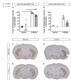
Helicobacter suis (H. suis)-infected mice are partially protected against the loss of dopaminergic neurons induced by intrastriatal 6-hydroxydopamine (6-OHDA) injection. (A,B) Loss of dopaminergic neuron fiber density in the left compared to the right striatum based on tyrosine hydroxylase (TH) staining, 7 days after intrastriatal injection with either 6-OHDA (n = 2–5) or vehicle (n = 1–6), of mice infected with H. suis (black) or the control broth (white) for a short- (A) or long-term period (B). (CF) Representative whole-brain section images of the tyrosine hydroxylase staining (brown) for dopaminergic neuron fiber density in the striatum of mice from a control/vehicle subgroup (C), H. suis/vehicle subgroup (D), control/6-OHDA subgroup (E), and H. suis/6-OHDA subgroup (F). **** p < 0.0001.
Figure 6
Figure 6
6-hydroxydopamine (6-OHDA) induced gliosis is not influenced by short-term Helicobacter suis (H. suis) infection. (A) Percentage (%) of IBA1-positive microglial cells more in the left injected striatum compared to the right non-injected striatum based on manual analysis, 7 days after intrastriatal injection with either 6-OHDA (n = 3–5) or vehicle (n = 5–6), of mice infected with H. suis (black) or the control broth (white) for a short-term period. (B) Percentage (%) of GFAP-positive astrocytes more in the left injected striatum compared to the right non-injected striatum based on automated analysis, 7 days after intrastriatal injection with either 6-OHDA (n = 3–5) or vehicle (n = 5–6), of mice infected with H. suis (black) or the control broth (white) for a short-term period. * 0.01 ≤ p < 0.05; ** 0.001 ≤ p < 0.01.
Figure 7
Figure 7
Helicobacter suis (H. suis) infection is associated with an increased gene expression of endogenous peroxidases. (AF) Relative mRNA gene expression of the endogenous peroxidases super oxide dismutase 2 (Sod2) (A,B), catalase (Cat) (C,D), and glutathione peroxidase 2 (Gpx2) (E,F) in the forebrain, 7 days after intrastriatal injection with either 6-hydroxydopamine (6-OHDA) (n = 5) or vehicle (n = 4–5), of mice infected with H. suis (black) or the control broth (white) for a short-term period (A,C,E) or a long-term period (B,D,F). * 0.01 ≤ p < 0.05; *** 0.0001 ≤ p < 0.001.
Figure 8
Figure 8
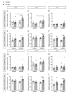
Helicobacter suis (H. suis) infection is associated with an increased gene expression of antioxidant nuclear factor (erythroid-derived 2)-like 2 (Nrf2)-associated genes. (AL) Relative mRNA gene expression of Nrf2 (A,B) and its downstream regulators glutamate-cysteine ligase modifier subunit (Gclm) (C,D), glutamate-cysteine ligase catalytic subunit (Gclc) (E,F), glutathi-one-disulfide reductase (Gsr) (G,H), heme oxygenase 1 (Hmox1) (I,J), and NADPH oxidase 2 (Nox2) (K,L) in the forebrain, 7 days after intrastriatal injection with either 6-hydroxydopamine (6-OHDA) (n = 5) or vehicle (n = 4–5), of mice infected with H. suis (black) or the control broth (white) for a short-term period (A,C,E,G,I,K) or a long-term period (B,D,F,H,J,L). * 0.01 ≤ p < 0.05; ** 0.001 ≤ p < 0.01; *** 0.0001 ≤ p < 0.001.

Similar articles

Cited by

References

    1. Collaborators G., Dorsey R.E., Elbaz A., Nichols E., Abd-Allah F., Abdelalim A., Adsuar J.C., Ansha M., Brayne C., Choi J.-Y.J., et al. Global, Regional, and National Burden of Parkinson’s Disease, 1990–2016: A Systematic Analysis for the Global Burden of Disease Study 2016. Lancet Neurol. 2018;17:939–953. doi: 10.1016/s1474-4422(18)30295-3. - DOI - PMC - PubMed
    1. Dorsey R.E., Sherer T., Okun M.S., Bloem B.R. The Emerging Evidence of the Parkinson Pandemic. J. Parkinson’s Dis. 2018;8:S3–S8. doi: 10.3233/JPD-181474. - DOI - PMC - PubMed
    1. Wirdefeldt K., Adami H.-O., Cole P., Trichopoulos D., Mandel J. Epidemiology and Etiology of Parkinson’s Disease: A Review of the Evidence. Eur. J. Epidemiol. 2011;26:1. doi: 10.1007/s10654-011-9581-6. - DOI - PubMed
    1. Johnson M.E., Stecher B., Labrie V., Brundin L., Brundin P. Triggers, Facilitators, and Aggravators: Redefining Parkinson’s Disease Pathogenesis. Trends Neurosci. 2018;42:4–13. doi: 10.1016/j.tins.2018.09.007. - DOI - PMC - PubMed
    1. Poewe W., Seppi K., Tanner C.M., Halliday G.M., Brundin P., Volkmann J., Schrag A.-E.E., Lang A.E. Parkinson Disease. Nature reviews. Dis. Primers. 2017;3:17013. doi: 10.1038/nrdp.2017.13. - DOI - PubMed

MeSH terms