Review
. 2021 Oct 20;22(21):11338.
doi: 10.3390/ijms222111338.

Mitochondria as a Cellular Hub in Infection and Inflammation

Affiliations
Review

Mitochondria as a Cellular Hub in Infection and Inflammation

Pauline Andrieux et al. Int J Mol Sci. .

Abstract

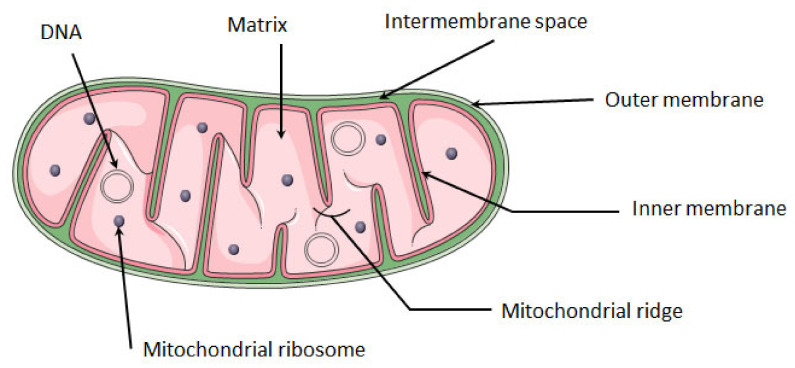
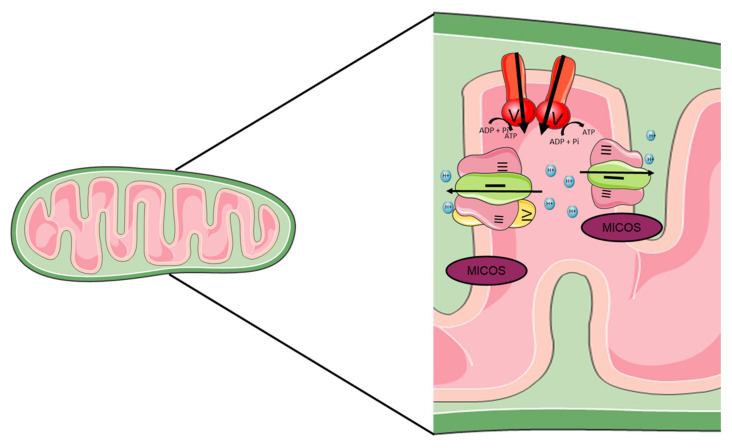
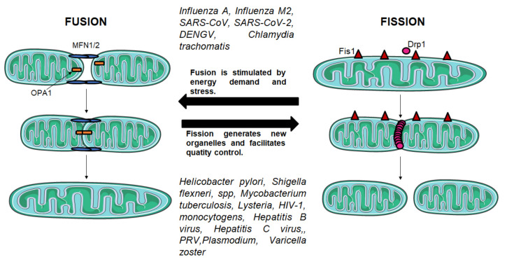
Mitochondria are the energy center of the cell. They are found in the cell cytoplasm as dynamic networks where they adapt energy production based on the cell's needs. They are also at the center of the proinflammatory response and have essential roles in the response against pathogenic infections. Mitochondria are a major site for production of Reactive Oxygen Species (ROS; or free radicals), which are essential to fight infection. However, excessive and uncontrolled production can become deleterious to the cell, leading to mitochondrial and tissue damage. Pathogens exploit the role of mitochondria during infection by affecting the oxidative phosphorylation mechanism (OXPHOS), mitochondrial network and disrupting the communication between the nucleus and the mitochondria. The role of mitochondria in these biological processes makes these organelle good targets for the development of therapeutic strategies. In this review, we presented a summary of the endosymbiotic origin of mitochondria and their involvement in the pathogen response, as well as the potential promising mitochondrial targets for the fight against infectious diseases and chronic inflammatory diseases.

Keywords: infection; infection disease; inflammation; inflammatory disease; mitochondria; mitochondria dysfunction; mitochondrial bioenergetics.


Conflict of interest statement

The authors declare no conflict of interest.

Figures

Figure 1
Figure 1
Structure of mitochondria. The mitochondrion is composed of a double membrane: the inner membrane and the outer membrane. Between these membranes is the intra-membrane space. The inner membrane forms invaginations called ridges where the OXPHOS complexes are located. The mitochondrial matrix contains several copies of mitochondrial circular DNA and ribosomes. Mitochondria image adapted from Smart. Available online: https://smart.servier.com (accessed on 10 August 2021).
Figure 2
Figure 2
Mitochondrial respiratory chain complex. The mitochondrial respiratory chain is located at the inner membrane of the mitochondria. Composed of four complexes and two coenzymes, it allows the production of ATP through oxidative phosphorylation. Complex I (NADH: coenzyme Q oxidoreductase) and II (succinate dehydrogenase) will each transfer two electrons to the quinone coenzyme Q10 (CoQ10). The two electrons transferred from complex I come from the oxidation of NADH, and those from complex II come from oxidation of succinate to fumarate. CoQ10 will allow the transfer of electrons to complex III (CoQ10-cytochrome C oxidoreductase). The complex III will then pass these electrons to the cytochrome C, which makes the link with the complex IV (cytochrome C oxidase). The complex IV reduces O2 in a H2O molecule. Complexes I, III and IV are proton pumps, which allow the passage of protons from the matrix to the intermembrane space, in the opposite direction of the gradient. Complexes I and III allow the passage of four protons and complex IV of two protons. Once the intermembrane space is enriched with protons, the last complex of the chain, ATP synthase will allow the passage of protons in the direction of the gradient. This proton flow will allow the synthesis of ATP from ADP.
Figure 3
Figure 3
Structure of mitochondrial supercomplexes. The structure of the mitochondrial ridges is very important in the function of mitochondria and in particular in the stability of the super complex. Several proteins are involved in the stability of these ridges, the ATP synthase which will be at the top and the mitochondrial contact site and cristae organizing system (MICOS) which will be at the bottom. Between the two will be the super complexes. The two main super-complexes found in mammals I + III2 + IV1–4 and I + III2 are shown.
Figure 4
Figure 4
Fusion and fission mechanisms. Fusion and fission mechanisms are important in the regulation of mitochondrial morphology and function. Fission is mediated by DRP1, a GTPase which is recruited from the cytosol by FIS1 to form spirals around mitochondria that constrict both inner and outer membranes. Fusion between mitochondrial outer membranes is mediated by MFN1 and MFN2, whereas fusion between mitochondrial inner membranes is mediated by OPA1.
Figure 5
Figure 5
The mitochondria at the center of the RLR, NLR and TLR pathways. The mitochondrion plays an important role in the innate immune response, as it participates in the main pathways involved in the immune response: the TLR, RLR and NLR pathways. The TLR pathway is involved in the antimicrobial response. PAMPs will activate the TLRs. The membrane-spanning TLRs (TLR1, TLR2, TLR4) and endosome-bonded TLRs (TLR3, TLR7, TLR8 and TLR9) are activated and trigger the expression of pro-inflammatory cytokines via the NF-kB pathway. The TLRs activate the kinases Mst1 and Mst2, which activate the GTPase Rac allowing the assembly of the TRAF6-ECSIT complex. The TRAF6 translocates to the mitochondria and binds to ECSIT, forming the TRAF6-ECSIT complex, which lead to an increase in mitochondrial ROS (mtROS) production as well as the proximity of the mitochondria and the phagosome. The bacteria in the phagosome will be destroyed by the action of mtROS. The RLR pathway is involved in antiviral response. Different PAMPs such as dsRNA, ssRNA and 5′-triphosphate (ppp) will activate two cytoplasmic PRRs, RIG-1 and MDA5. These PRR are regulated by DHX58, where DHX58 positively regulates MDA5 and negatively regulates RIG-1. After the interaction with DHX58/RIG1/MDA5, MAVS is activated by recruiting different proteins, such as TRAF3 and TRAF6. The interaction between MAVS and the MFN1 and MFN2 fusion proteins (MFNs) are essential for RLR pathway signaling. TRAF3 will activate the TANK/NEMO/iKKe/TBK complex, which will phosphorylate IRF7 and IRF3 dimers, allowing their translocation into the nucleus and their binding to ISRE sequences at the promoters of type I IFN-regulated genes, while TRAF6 will activate the IKK complex. This complex activates NF-ĸB by phosphorylating its inhibitor IKBα. Once activated, NF-ĸB translocates to the nucleus to induce the expression of pro-inflammatory genes. The NLR pathway is involved in a mechanism for fighting infection and cellular damage. The main complex of this pathway is the NLRP3 inflammasome. The inflammasome is a multiprotein complex containing ASC, pro-caspase 1 and NLRP3. The activation of NLRP3 inflammasome is done both on transcriptional and post translational levels. The first is related to TLR activation and production of pro-IL1b and pro-IL18. The second signal is based on the detection of PAMPs and DAMPs that leads to NLRP3 oligomerization and interaction with ASC. Then, ASC recruits pro-caspase 1 via the CARD domain. Once caspase 1 is activated, it cleaves the pro-IL1b and pro-IL18 which will allow the release of these inflammatory cytokines in the extracellular milieu. In addition, caspase 1 cleaves Gsdmd, which will allow pore creation at the plasma membrane and pyroptosis. In addition, MAVS is also necessary for the recruitment of NLRP3 in mitochondrial membranes. Cardiolipin and mtROS promote the activation of NLRP3 inflammasome.

References

    1. Mileykovskaya E., Dowhan W. Cardiolipin membrane domains in prokaryotes and eukaryotes. Biochim. Biophys. Acta (BBA) Biomembr. 2009;1788:2084–2091. doi: 10.1016/j.bbamem.2009.04.003. - DOI - PMC - PubMed
    1. Nass S., Nass M.M. Intramitochondrial Fibers with DNA Characteristics. Ii. Enzymatic and Other Hydrolytic Treatments. J. Cell Biol. 1963;19:613–629. doi: 10.1083/jcb.19.3.613. - DOI - PMC - PubMed
    1. Sagan L. On the origin of mitosing cells. J. Theor. Biol. 1967;14:225–274. doi: 10.1016/0022-5193(67)90079-3. - DOI - PubMed
    1. Martin W., Müller M. The hydrogen hypothesis for the first eukaryote. Nature. 1998;392:37–41. doi: 10.1038/32096. - DOI - PubMed
    1. Gould S.B., Garg S.G., Martin W.F. Bacterial Vesicle Secretion and the Evolutionary Origin of the Eukaryotic Endomembrane System. Trends Microbiol. 2016;24:525–534. doi: 10.1016/j.tim.2016.03.005. - DOI - PubMed

LinkOut - more resources