. 2021 Oct 21;22(21):11368.
doi: 10.3390/ijms222111368.

PRC1 Stabilizes Cardiac Contraction by Regulating Cardiac Sarcomere Assembly and Cardiac Conduction System Construction

Affiliations

PRC1 Stabilizes Cardiac Contraction by Regulating Cardiac Sarcomere Assembly and Cardiac Conduction System Construction

Xixia Peng et al. Int J Mol Sci. .

Abstract

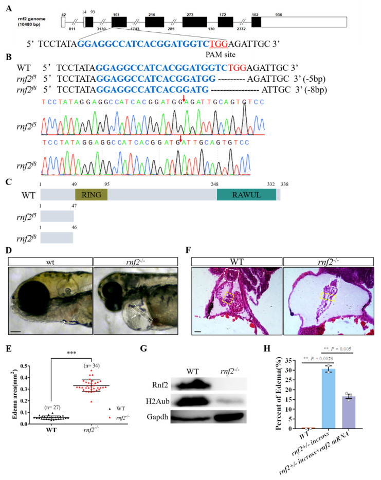
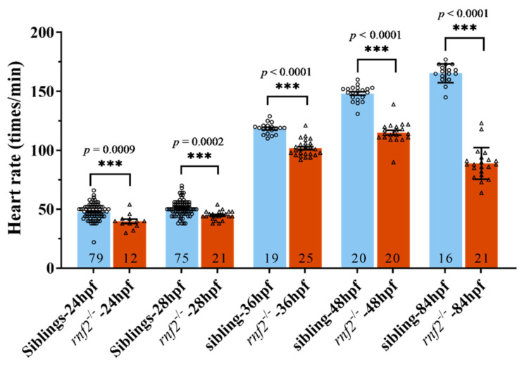
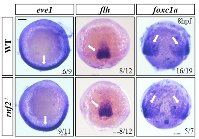
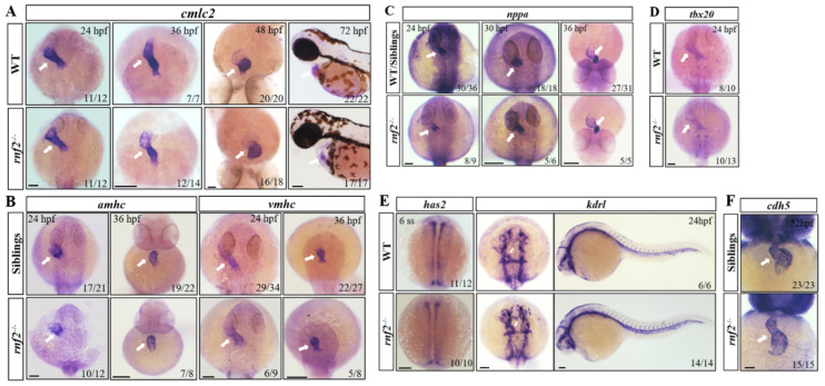
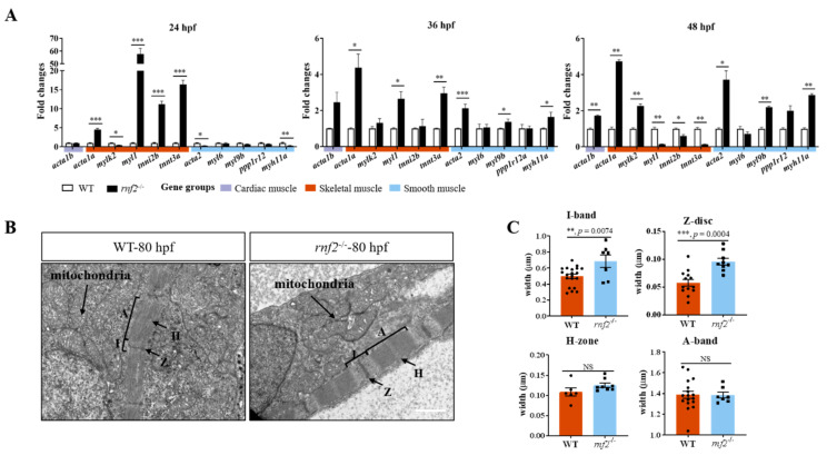
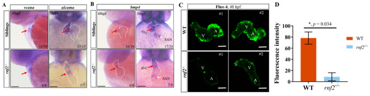
Cardiac development is a complex process that is strictly controlled by various factors, including PcG protein complexes. Several studies have reported the critical role of PRC2 in cardiogenesis. However, little is known about the regulation mechanism of PRC1 in embryonic heart development. To gain more insight into the mechanistic role of PRC1 in cardiogenesis, we generated a PRC1 loss-of-function zebrafish line by using the CRISPR/Cas9 system targeting rnf2, a gene encoding the core subunit shared by all PRC1 subfamilies. Our results revealed that Rnf2 is not involved in cardiomyocyte differentiation and heart tube formation, but that it is crucial to maintaining regular cardiac contraction. Further analysis suggested that Rnf2 loss-of-function disrupted cardiac sarcomere assembly through the ectopic activation of non-cardiac sarcomere genes in the developing heart. Meanwhile, Rnf2 deficiency disrupts the construction of the atrioventricular canal and the sinoatrial node by modulating the expression of bmp4 and other atrioventricular canal marker genes, leading to an impaired cardiac conduction system. The disorganized cardiac sarcomere and defective cardiac conduction system together contribute to defective cardiac contraction. Our results emphasize the critical role of PRC1 in the cardiac development.

Keywords: PRC1; Rnf2; cardiac conduction system; cardiac contraction; sarcomere assembly.


Conflict of interest statement

The authors declare no conflict of interest.

Figures

Figure 1
Figure 1
Rnf2-null zebrafish mutant displayed severe cardiac defects. (A) The target site (sequence highlighted in blue) is located in exon 3. The PAM site is underlined and highlighted in red; (B) DNA sequencing identified that two mutant alleles carried a 5 bp (rnf2f5) and 8 bp (rnf2f8) deletion respectively. The deletion sites are indicated by red arrows; (C) Schematic diagram of the wild-type and mutant Rnf2 proteins. Zebrafish wild-type Rnf2 contained an N-terminal Ring-finger domain (yellow) and a C-terminal RAWUL domain. The two mutant proteins were truncated before the RING-finger domain; (D) The rnf2−/− larvae displayed severe pericardia edema. The hearts are indicated by white arrows, the stringy heart in rnf2−/− is depicted by white dotted lines, scale bar: 0.2 mm; (E) Scatter plot showing the sectional area of edema in wild-type (black) and rnf2−/− (red). (F) Histological sections of 4 dpf heart, boxes in yellow indicate the AVC and valves, white boxes indicate the bulbous arteriosus, scale bar: 50 μm, n(WT) = 2, n (rnf2−/−) = 3; (G) Western blot verified the deletion of Rnf2 protein. Gapdh was set as internal reference; (H) Bar graph showing the percentage of embryos with edema. **, p < 0.01; ***, p < 0.001; n, sample number.
Figure 2
Figure 2
The cardiac contraction was disrupted in Rnf2-null zebrafish embryos. Scatter plot showing the heart rates of wild-type (blue) and rnf2−/− (red) embryos at different developmental stages. Numbers in the bottoms of the bars indicate the sample number of each group. ***, p < 0.001.
Figure 3
Figure 3
The mesoderm formed normally in Rnf2-null zebrafish embryos. WISH results showing expression of mesoderm markers eve1 (ventral mesoderm), flh (axial mesoderm), and foxc1a (paraxial mesoderm). The expression regions are indicated by white arrows. Scale bar: 0.2 mm.
Figure 4
Figure 4
Expression of cardiomyocyte markers examined by in situ hybridization. (A) cmlc2 at different developmental stages; (B) Atrial (amhc) and ventricle (vmhc) cardiomyocyte markers at 24 hpf and 36 hpf; (C) Growth factor nppa at 24 hpf, 30 hpf, and 36 hpf; (D) tbx20 at 24 hpf; (E) Expression of endocardial precursor marker gene has2 and endothelial marker gene kdrl in the heart and blood vessels, white arrows indicate the heart tube; (F) Expression of endothelial marker gene cdh5 in the heart. The expression of genes in the heart is indicated by white arrows. Scale bar: 0.2 mm.
Figure 5
Figure 5
Rnf2 deficiency disorganized sarcomere assembly in zebrafish hearts. (A) Real-time PCR tested the expression of skeletal and smooth muscle genes at 36 hpf (left) and 48 hpf (right). The fold changes of relative mRNA levels are presented as mean ± SEM. The expression in wild-types was normalized to 1. The experiment was repeated on three separate occasions. n = 159, 111, and 161 for rnf2−/− group at 24 hpf, 36 hpf, and 48 hpf, respectively; n = 145, 113, and 164 for WT group at 24 hpf, 36 hpf, and 48 hpf, respectively. (B) Cardiac TEM revealed the sarcomere of cardiac muscle was abnormal in rnf2−/− hearts. A, A-band; I, I-band; H, H-zone; Z, Z-disc. Scale bar: 1.0 μm. n = 3. (C) Bar graph showing the width of A-band, I-band, Z-disc, and H-zone in wild-type and rnf2−/− cardiac sarcomeres. *, p < 0.05; **, p < 0.01; ***, p < 0.001; NS, no significant.
Figure 6
Figure 6
Rnf2 deficiency caused defects in the cardiac conduction system. (A) Expression of AVC myocardial markers vcana and alcama; (B) Expression of AVC endocardial and SAN marker bmp4. Red arrows indicate AVC; (C) Fluo-4 staining detects calcium signal intensity in the hearts of rnf2−/− or wild-type embryos; V, ventricle; A, Atrium; #1/#2, sample number; n = 2. (D) Quantification of fluorescence intensity in Figure 6C. *, p < 0.05; Scale bar: 0.2 mm.

Similar articles

Cited by

References

    1. Bakkers J., Verhoeven M.C., Abdelilah-Seyfried S. Shaping the zebrafish heart: From left-right axis specification to epithelial tissue morphogenesis. Dev. Biol. 2009;330:213–220. doi: 10.1016/j.ydbio.2009.04.011. - DOI - PubMed
    1. Houk A.R., Yelon D. Strategies for analyzing cardiac phenotypes in the zebrafish embryo. In: Detrich H.W., Westerfield M., Zon L.I., editors. Zebrafish: Cellular and Developmental Biology, Pt B: Developmental Biology. Elsevier; Amsterdam, The Netherlands: 2016. pp. 335–368. - PMC - PubMed
    1. Miura G.I., Yelon D. A guide to analysis of cardiac phenotypes in the zebrafish embryo. Methods Cell Biol. 2011;101:161–180. - PMC - PubMed
    1. Peterson R.T., Mably J.D., Chen J.-N., Fishman M.C. Convergence of distinct pathways to heart patterning revealed by the small molecule concentramide and the mutation heart-and-soul. Curr. Biol. 2001;11:1481–1491. doi: 10.1016/S0960-9822(01)00482-1. - DOI - PubMed
    1. Yelon D., Horne S.A., Stainier D.Y.R. Restricted expression of cardiac myosin genes reveals regulated aspects of heart tube assembly in zebrafish. Dev. Biol. 1999;214:23–37. doi: 10.1006/dbio.1999.9406. - DOI - PubMed

MeSH terms

Substances

LinkOut - more resources