Review
. 2021 Oct 21;22(21):11377.
doi: 10.3390/ijms222111377.

Common Pathogenic Mechanisms in Centronuclear and Myotubular Myopathies and Latest Treatment Advances

Affiliations
Review

Common Pathogenic Mechanisms in Centronuclear and Myotubular Myopathies and Latest Treatment Advances

Raquel Gómez-Oca et al. Int J Mol Sci. .

Abstract

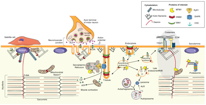
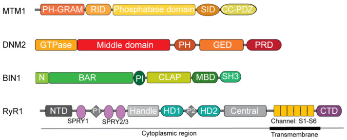
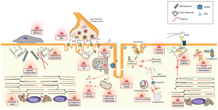
Centronuclear myopathies (CNM) are rare congenital disorders characterized by muscle weakness and structural defects including fiber hypotrophy and organelle mispositioning. The main CNM forms are caused by mutations in: the MTM1 gene encoding the phosphoinositide phosphatase myotubularin (myotubular myopathy), the DNM2 gene encoding the mechanoenzyme dynamin 2, the BIN1 gene encoding the membrane curvature sensing amphiphysin 2, and the RYR1 gene encoding the skeletal muscle calcium release channel/ryanodine receptor. MTM1, BIN1, and DNM2 proteins are involved in membrane remodeling and trafficking, while RyR1 directly regulates excitation-contraction coupling (ECC). Several CNM animal models have been generated or identified, which confirm shared pathological anomalies in T-tubule remodeling, ECC, organelle mispositioning, protein homeostasis, neuromuscular junction, and muscle regeneration. Dynamin 2 plays a crucial role in CNM physiopathology and has been validated as a common therapeutic target for three CNM forms. Indeed, the promising results in preclinical models set up the basis for ongoing clinical trials. Another two clinical trials to treat myotubular myopathy by MTM1 gene therapy or tamoxifen repurposing are also ongoing. Here, we review the contribution of the different CNM models to understanding physiopathology and therapy development with a focus on the commonly dysregulated pathways and current therapeutic targets.

Keywords: amphiphysin; autophagy; centronuclear myopathy; dynamin; membrane trafficking; myotubular myopathy; myotubularin; ryanodine receptor; satellite cell; triads.


Conflict of interest statement

B.S. Cowling and J. Laporte are co-founders of Dynacure. R. Gomez-Oca and B.S. Cowling are employees of Dynacure.

Figures

Figure 1
Figure 1
Schematic illustration of localization and functions of MTM1, DNM2, BIN1, and RyR1 in skeletal muscle. ECM (extracellular matrix), ALR (autophagic lysosome reformation), Ub (ubiquitin), Ach (acetylcholine), MVB (multivesicular bodies). The figure uses modified images from Servier Medical Art Commons Attribution 3.0 Unported License (http://smart.servier.com, accessed on 29 August 2021).
Figure 2
Figure 2
Scheme of MTM1, DNM2, BIN1, and RyR1 protein domains. MTM1: PH-GRAM (Pleckstrin homology-glucosyltransferase; Rab-like GTPase activator and myotubularin), RID (Rac1-induced recruitment domain), SID (SET interacting domain), CC-PDZ (coiled coil; PSD95, disc large, ZO-1). DNM2: PH (Pleckstrin-homology), GED (GTPase effector domain), PRD (proline-rich domain). BIN1: N (N-terminal amphipathic helix α), BAR (Bin-amphiphysin-Rvs), PI (phosphoinositides), CLAP (clathrin and AP2 binding), MBD (Myc binding domain), SH3 (SRC homology 3). RyR1: NTD (N-terminal domain), SPRY (SPla and the RYanodine receptor), HD (helical domain), CTD (C-terminal domain).
Figure 3
Figure 3
Schematic illustration of pathogenic alterations in skeletal muscle caused by mutations in MTM1, BIN1, DNM2, and RYR1. ECM (extracellular matrix), ALR (autophagic lysosome reformation), SR (sarcoplasmic reticulum), Ub (ubiquitin), NMJ (neuromuscular junction), ECC (excitation-contraction coupling), Ach (acetylcholine), MVB (multivesicular bodies). The figure uses modified images from Servier Medical Art Commons Attribution 3.0 Unported License (http://smart.servier.com, accessed on 29 August 2021).

References

    1. Romero N.B. Centronuclear myopathies: A widening concept. Neuromuscul. Disord. 2010;20:223–228. doi: 10.1016/j.nmd.2010.01.014. - DOI - PubMed
    1. Spiro A.J., Shy G.M., Gonatas N.K. Myotubular myopathy. Persistence of fetal muscle in an adolescent boy. Arch. Neurol. 1966;14:1–14. doi: 10.1001/archneur.1966.00470070005001. - DOI - PubMed
    1. Laporte J., Hu L.J., Kretz C., Mandel J.L., Kioschis P., Coy J.F., Klauck S.M., Poustka A., Dahl N. A gene mutated in X-linked myotubular myopathy defines a new putative tyrosine phosphatase family conserved in yeast. Nat. Genet. 1996;13:175–182. doi: 10.1038/ng0696-175. - DOI - PubMed
    1. Bitoun M., Maugenre S., Jeannet P.Y., Lacene E., Ferrer X., Laforet P., Martin J.J., Laporte J., Lochmuller H., Beggs A.H., et al. Mutations in dynamin 2 cause dominant centronuclear myopathy. Nat. Genet. 2005;37:1207–1209. doi: 10.1038/ng1657. - DOI - PubMed
    1. Bohm J., Biancalana V., Malfatti E., Dondaine N., Koch C., Vasli N., Kress W., Strittmatter M., Taratuto A.L., Gonorazky H., et al. Adult-onset autosomal dominant centronuclear myopathy due to BIN1 mutations. Brain. 2014;137:3160–3170. doi: 10.1093/brain/awu272. - DOI - PubMed

MeSH terms

LinkOut - more resources