

Ultramicronized Palmitoylethanolamide in the Management of Sepsis-Induced Coagulopathy and Disseminated Intravascular Coagulation
- PMID: 34768820
- PMCID: PMC8583705
- DOI: 10.3390/ijms222111388
Ultramicronized Palmitoylethanolamide in the Management of Sepsis-Induced Coagulopathy and Disseminated Intravascular Coagulation
Abstract
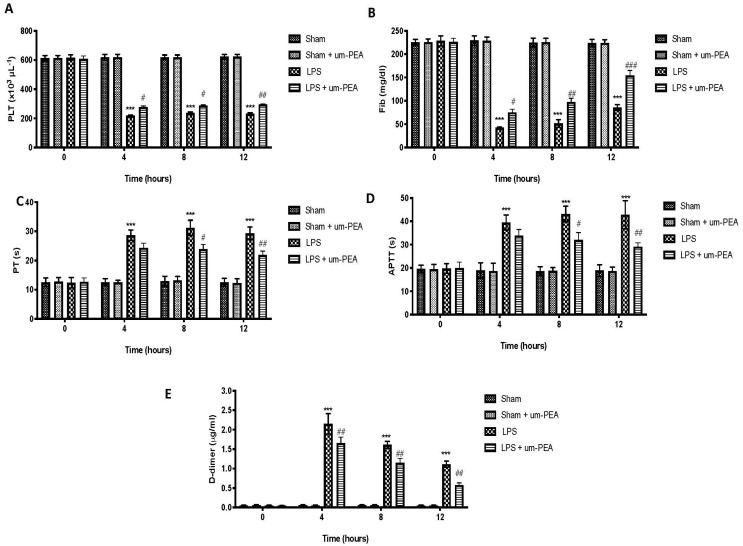
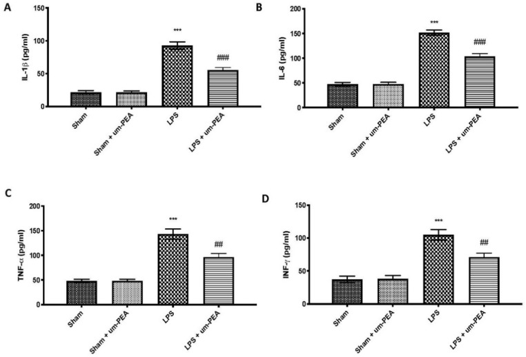
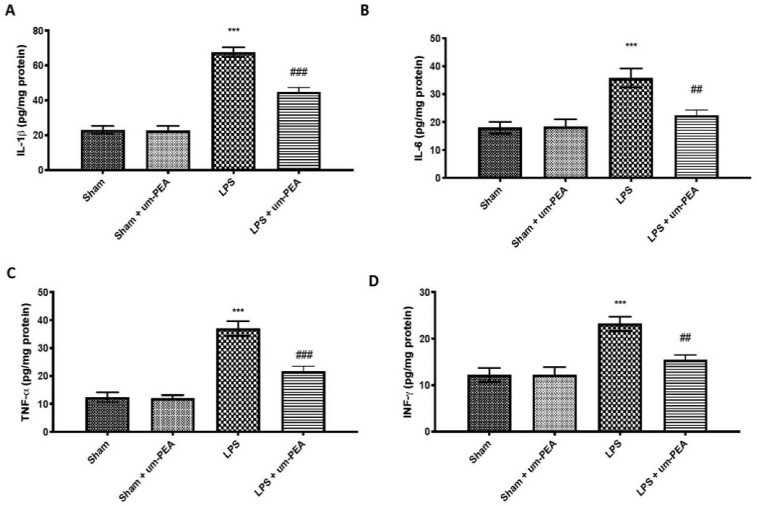
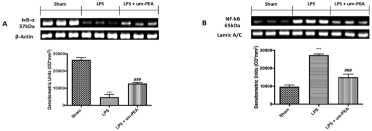
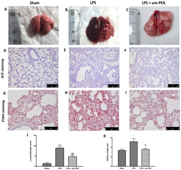
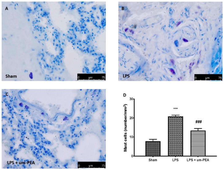
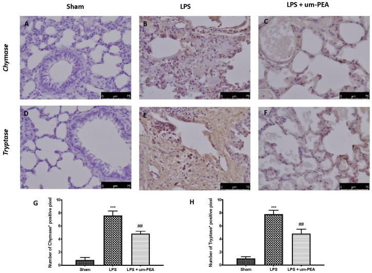
Disseminated intravascular coagulation (DIC) is a severe condition characterized by the systemic formation of microthrombi complicated with bleeding tendency and organ dysfunction. In the last years, it represents one of the most frequent consequences of coronavirus disease 2019 (COVID-19). The pathogenesis of DIC is complex, with cross-talk between the coagulant and inflammatory pathways. The objective of this study is to investigate the anti-inflammatory action of ultramicronized palmitoylethanolamide (um-PEA) in a lipopolysaccharide (LPS)-induced DIC model in rats. Experimental DIC was induced by continual infusion of LPS (30 mg/kg) for 4 h through the tail vein. Um-PEA (30 mg/kg) was given orally 30 min before and 1 h after the start of intravenous infusion of LPS. Results showed that um-PEA reduced alteration of coagulation markers, as well as proinflammatory cytokine release in plasma and lung samples, induced by LPS infusion. Furthermore, um-PEA also has the effect of preventing the formation of fibrin deposition and lung damage. Moreover, um-PEA was able to reduce the number of mast cells (MCs) and the release of its serine proteases, which are also necessary for SARS-CoV-2 infection. These results suggest that um-PEA could be considered as a potential therapeutic approach in the management of DIC and in clinical implications associated to coagulopathy and lung dysfunction, such as COVID-19.
Keywords: coagulation; disseminated intravascular coagulation; inflammation; sepsis; ultramicronized palmitoylethanolamide.
Conflict of interest statement
Salvatore Cuzzocrea is a coinventor on patent WO2013121449 A8 (Epitech Group Srl), which deals with methods and compositions for the modulation of amidases capable of hydrolyzing N-acylethanolamines employable in the treatment of inflammatory diseases. This invention is wholly unrelated to the present study. Moreover, Cuzzocrea is also, with Epitech Group, a coinventor on the patents EP 2 821 083, MI2014 A001495, and 102015000067344, which are unrelated to the study. The remaining authors report no conflicts of interest.
Figures
Similar articles
-
Management of Acute Lung Injury: Palmitoylethanolamide as a New Approach.Int J Mol Sci. 2021 May 24;22(11):5533. doi: 10.3390/ijms22115533. Int J Mol Sci. 2021. PMID: 34073872 Free PMC article.
-
Coagulopathy in COVID-19.J Thromb Haemost. 2020 Sep;18(9):2103-2109. doi: 10.1111/jth.14975. Epub 2020 Jul 21. J Thromb Haemost. 2020. PMID: 32558075 Free PMC article. Review.
-
Micronized/ultramicronized palmitoylethanolamide displays superior oral efficacy compared to nonmicronized palmitoylethanolamide in a rat model of inflammatory pain.J Neuroinflammation. 2014 Aug 28;11:136. doi: 10.1186/s12974-014-0136-0. J Neuroinflammation. 2014. PMID: 25164769 Free PMC article.
-
Effect of Ultra-Micronized-Palmitoylethanolamide and Acetyl-l-Carnitine on Experimental Model of Inflammatory Pain.Int J Mol Sci. 2021 Feb 17;22(4):1967. doi: 10.3390/ijms22041967. Int J Mol Sci. 2021. PMID: 33671213 Free PMC article.
-
Coagulopathy of sepsis.Thromb Haemost. 2004 Feb;91(2):213-24. doi: 10.1160/TH03-03-0182. Thromb Haemost. 2004. PMID: 14961146 Review.
Cited by
-
Comparison of the prognostic value of four different critical illness scores in patients with sepsis-induced coagulopathy.Open Life Sci. 2023 Aug 9;18(1):20220659. doi: 10.1515/biol-2022-0659. eCollection 2023. Open Life Sci. 2023. PMID: 37588996 Free PMC article.
-
From Molecular Mechanisms to Clinical Therapy: Understanding Sepsis-Induced Multiple Organ Dysfunction.Int J Mol Sci. 2024 Jul 16;25(14):7770. doi: 10.3390/ijms25147770. Int J Mol Sci. 2024. PMID: 39063011 Free PMC article. Review.
-
Micro-inflammation related gene signatures are associated with clinical features and immune status of fibromyalgia.J Transl Med. 2023 Sep 5;21(1):594. doi: 10.1186/s12967-023-04477-w. J Transl Med. 2023. PMID: 37670381 Free PMC article.
-
Role of neutrophil extracellular traps in inflammatory evolution in severe acute pancreatitis.Chin Med J (Engl). 2022 Dec 5;135(23):2773-2784. doi: 10.1097/CM9.0000000000002359. Chin Med J (Engl). 2022. PMID: 36729096 Free PMC article. Review.
-
Targeting Nrf2 and NF-κB Signaling Pathways in Inflammatory Pain: The Role of Polyphenols from Thinned Apples.Molecules. 2023 Jul 13;28(14):5376. doi: 10.3390/molecules28145376. Molecules. 2023. PMID: 37513248 Free PMC article.
References
MeSH terms
Substances
LinkOut - more resources
Full Text Sources
Other Literature Sources
Medical
Miscellaneous
