. 2021 Oct 26;22(21):11508.
doi: 10.3390/ijms222111508.

Structural and Functional Analysis of Female Sex Hormones against SARS-CoV-2 Cell Entry

Affiliations

Structural and Functional Analysis of Female Sex Hormones against SARS-CoV-2 Cell Entry

Jorge Alberto Aguilar-Pineda et al. Int J Mol Sci. .

Abstract

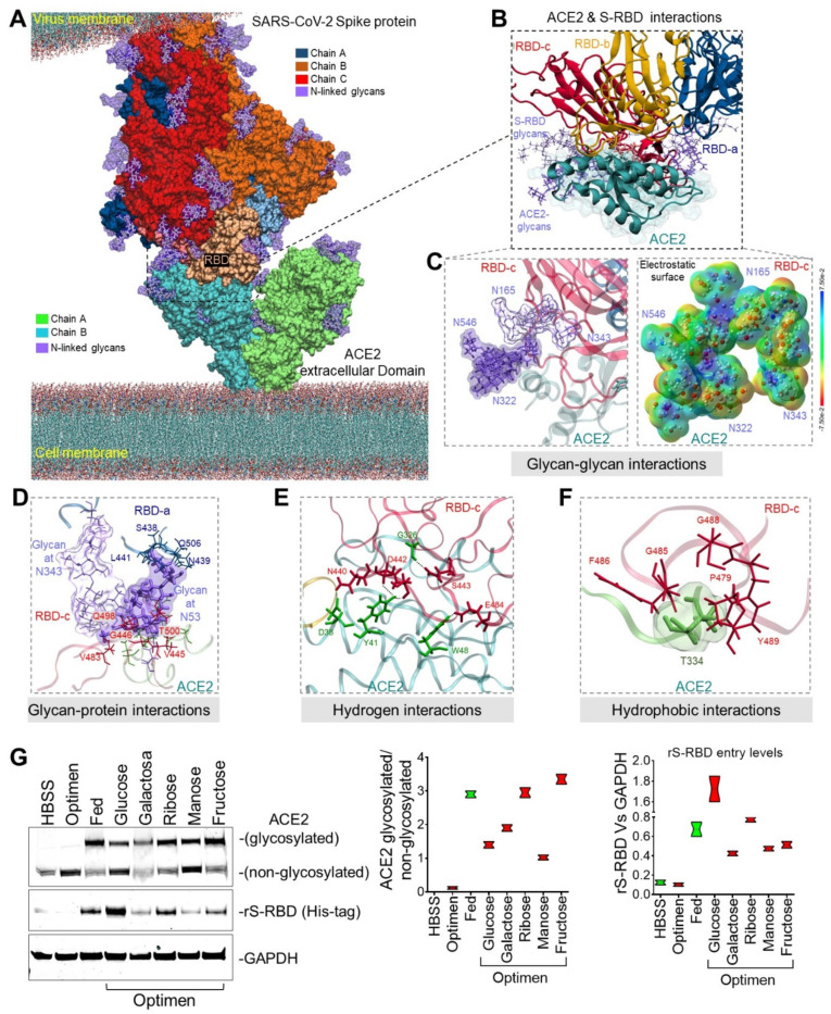
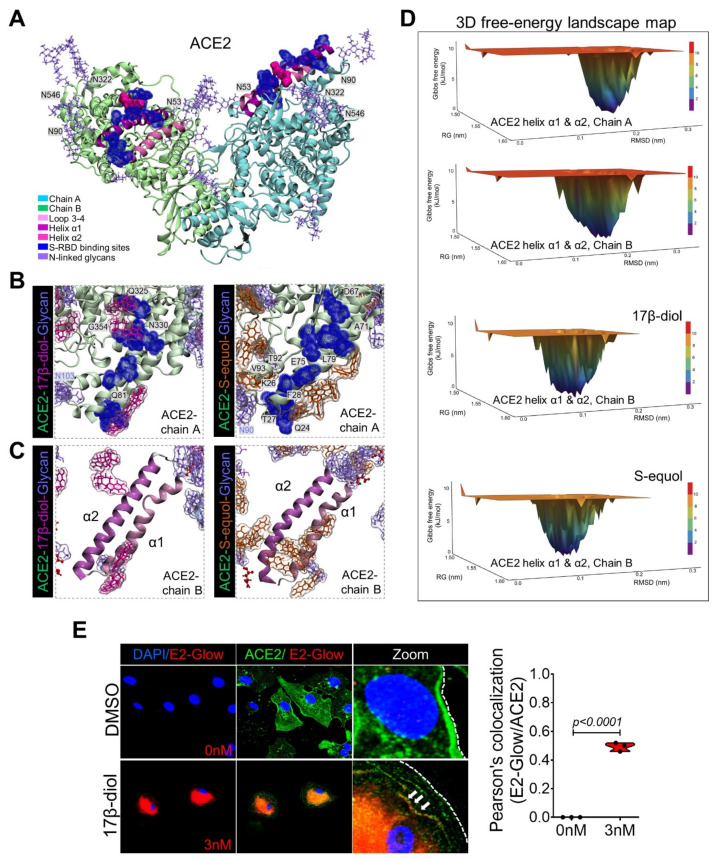
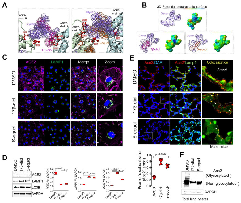
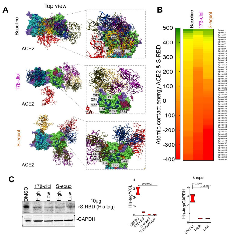
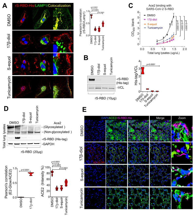
Emerging evidence suggests that males are more susceptible to severe infection by the SARS-CoV-2 virus than females. A variety of mechanisms may underlie the observed gender-related disparities including differences in sex hormones. However, the precise mechanisms by which female sex hormones may provide protection against SARS-CoV-2 infectivity remains unknown. Here we report new insights into the molecular basis of the interactions between the SARS-CoV-2 spike (S) protein and the human ACE2 receptor. We further report that glycosylation of the ACE2 receptor enhances SARS-CoV-2 infectivity. Importantly, estrogens can disrupt glycan-glycan interactions and glycan-protein interactions between the human ACE2 and the SARS-CoV-2 thereby blocking its entry into cells. In a mouse model of COVID-19, estrogens reduced ACE2 glycosylation and thereby alveolar uptake of the SARS-CoV-2 spike protein. These results shed light on a putative mechanism whereby female sex hormones may provide protection from developing severe infection and could inform the development of future therapies against COVID-19.

Keywords: ACE2; COVID-19; estrogenes; sex hormones.


Conflict of interest statement

The authors declare no conflict of interest.

Figures

Figure 1
Figure 1
Molecular bases of glycosylated hACE2 and SARS-CoV-2 Spike protein complex. (A) 3D membrane surface representation of glycosylated ACE2 in complex with the SARS-CoV-2 Spike protein. (B) Close up of the interacting environment between ACE2 and the S-RBD trimer. (C) The left panel demonstrates glycan–glycan interactions between ACE2 (dark purple surface) and S-RBDc (light purple surface). The right panel shows that glycan–glycan contacts do not affect their molecular electrostatic potentials (MEPs) properties. The energy scale ranges from −0.075 μa (red) to 0.075 μa (blue). (D) ACE2 glycan at N53 forms glycan–protein contact with residues on the S-RBDa and S-RBDc proteins. (E) The ACE2 glycosylation induces the formation of hydrogen bonds that engages the helix α1 in the binding with multiple residues on the S-RBDc. (F) Hydrophobic interactions occur between ACE2 at T334 and multiple residues on the S-RBDc. (G) Immunoblot shows the glycosylation status of the human ACE2 in HUVECs treated with different saccharides. Glucose-treated cells induced the greatest internalization of the recombinant S-RBD. Quantification of protein levels of three replicate experiments is shown. Student’s T-test, 2 tails. Bar graphs are presented as mean with error bars (±SD).
Figure 2
Figure 2
Estrogen effects on ACE2 structural energy. (A) 3D representation of the human ACE2 glycosylated residues and key regions used by the SARS-CoV-2 S protein to mediate entry into cells. S-RBD-binding sites are colored in dark blue and glycans in purple. (B) 3D molecular interactions between ACE2 and 17β-diol (magenta) or S-equol (orange) molecules obtained by 100 ns of molecular dynamics simulations (MDS). (C) A plain representation of solvated ACE2-helix α1 and α2 substructures by estrogen molecules. (D) FEL maps represent the conformational energy of helix α1 and α2 substructures with estrogen molecules during MDS (last 20 ns of MDS). The energy scale ranges from 12 kJ/mol (red) to 0 kJ/mol (blue). (E) Immunofluorescence microscopy analysis on HUVECs cells treated with or without conjugated 17β-diol (E2-Glow), shows colocalization (yellow) between ACE2 (green) and 17β-diol (red).
Figure 3
Figure 3
Estrogens bind to ACE2 glycans to promote its internalization. (A) Glycan–estrogen interactions stabilize ACE2 Glycan-residues at E57, N53, K341, and V339 (red color). (B) MEP maps show the electrostatic impact of estrogen molecules on the surface of ACE2 glycans. The energy scale ranges from −0.075 μa (red) to 0.075 μa (blue). (C) Immunofluorescence staining of human ACE2 (magenta) and the lysosome marker LAMP1 (green), shows loss of ACE2 membrane levels in HUVECs treated with 17β-diol or S-equol compared with the control group (DMSO). (D) Immunoblot shows decreased levels of total ACE2 protein which associates with increased endocytosis activity as evidenced by immunoblot for LC3b and LAMP1. (E) Histologic analysis of wild-type mouse lungs after 48 h of intratracheal instillation with 17β-diol or S-equol shows loss of Ace2 signal (red) on the membrane of alveoli cells. Estrogen-treated lungs show greater Ace2-Lamp1 colocalization (white arrows) indicating internalization of the receptor. (F) Immunoblot shows decreased levels of total and glycosylated Ace2 proteins in estrogen-treated lungs from male mice. Quantification of protein levels of three replicate experiments is shown. Student’s t-test, 2 tails. Bar graphs are presented as mean with error bars (±SD).
Figure 4
Figure 4
Estrogen’s impact on ACE2 and S-RBD interactions. (A) Top view of the 3D ACE2 surface interacting with 5 top-scored S-RBDs (top 1—blue, 2—red, 3—orange, 4—purple, and top 5—yellow). S-RBDs were scored based on shape complementarity principles. (B) Heatmap of atomic contact energy between ACE2 and 57 S-RBDs shows spontaneous energy structures from most favorable (green) to less favorable S-RBD structures (red). Energy scale ranging from 500 Kcal/mol to −500 Kcal/mol. (C) Immunoblot of isolated proteins from cultured HUVECs shows a 90% inhibition of S-RBD entry into cells in estrogen-treated cells. Quantification of protein levels of three replicate experiments is shown. Student’s T-test, 2 tails. Bar graphs are presented as mean with error bars (±SD).
Figure 5
Figure 5
Estrogens block SARS-CoV-2 S protein uptake in the respiratory tract in vivo. (A,B) Immunofluorescence analysis of S-RBD entry into HUVECs pretreated estrogens (17β-diol or S-equol) or Tunicamycin (glycosylation inhibitor) followed by treatment with 10μg/mL of recombinant S-RBD (red) demonstrate that all 3 treatments cells reduced entry of S-RBD entry into cells via a reduction in Ace2 internalization as shown by colocalization with LAMP1 (green) and immunoblot. (C) ELISA-based binding assay using lung protein lysates from male mice (K18-ACE2) treated with 17β-diol (0.3 μM) or S-equol (1μM) or Tunicamycin (1 mg/Kg) shows reduced SARS-CoV-2 S protein affinity for the Ace2 receptor. (D) Immunoblot shows the glycosylation levels of Ace2 from total lung lysate in mice treated with estrogens or Tunicamycin. (E) Immunofluorescence of Ace2 (green) and RBD (magenta) in lungs from lysates from male mice (K18-ACE2) treated with estrogens or Tunicamycin.

Update of

References

    1. Glinsky G.V. Tripartite combination of candidate pandemic mitigation agents: Vitamin D, quercetin, and estradiol manifest properties of medicinal agents for targeted mitigation of the COVID-19 pandemic defined by genomics-guided tracing of SARS-CoV-2 targets in human cells. Biomedicines. 2020;8:129. - PMC - PubMed
    1. Sama I., Ravera A., Santema B.T., Van Goor H., Ter Maaten J.M., Cleland J.G.F., Rienstra M., Friedrich A.W., Samani N.J., Ng L.L., et al. Circulating plasma concentrations of angiotensin-converting enzyme 2 in men and women with heart failure and effects of renin–angiotensin–aldosterone inhibitors. Eur. Heart J. 2020;41:1810–1817. doi: 10.1093/eurheartj/ehaa373. - DOI - PMC - PubMed
    1. Tian W., Jiang W., Yao J., Nicholson C.J., Li R., Sigurslid H., Wooster L., Rotter J.I., Guo X., Malhotra R. Predictors of mortality in hospitalized COVID-19 patients: A systematic review and meta-analysis. J. Med. Virol. 2020;92:1875–1883. doi: 10.1002/jmv.26050. - DOI - PMC - PubMed
    1. Gebhard C., Regitz-Zagrosek V., Neuhauser H.K., Morgan R., Klein S.L. Impact of sex and gender on COVID-19 outcomes in Europe. Biol. Sex Differ. 2020;11:1–13. doi: 10.1186/s13293-020-00304-9. - DOI - PMC - PubMed
    1. Peckham H., de Gruijter N.M., Raine C., Radziszewska A., Ciurtin C., Wedderburn L.R., Rosser E.C., Webb K., Deakin C.T. Male sex identified by global COVID-19 meta-analysis as a risk factor for death and ITU admission. Nat. Commun. 2020;11:6317. doi: 10.1038/s41467-020-19741-6. - DOI - PMC - PubMed

MeSH terms

LinkOut - more resources