. 2021 Oct 26;22(21):11536.
doi: 10.3390/ijms222111536.

Small RNA F6 Provides Mycobacterium smegmatis Entry into Dormancy

Affiliations

Small RNA F6 Provides Mycobacterium smegmatis Entry into Dormancy

Artem Grigorov et al. Int J Mol Sci. .

Abstract

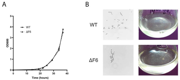
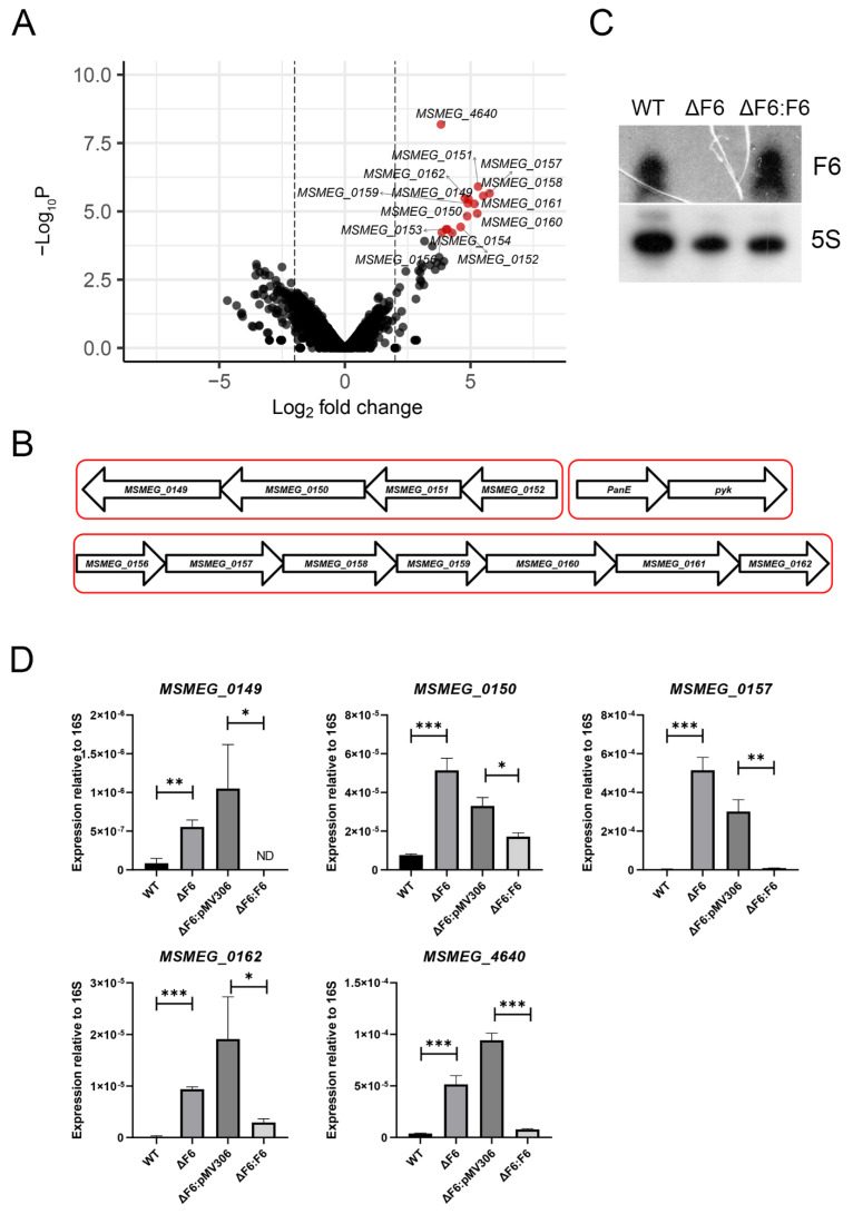
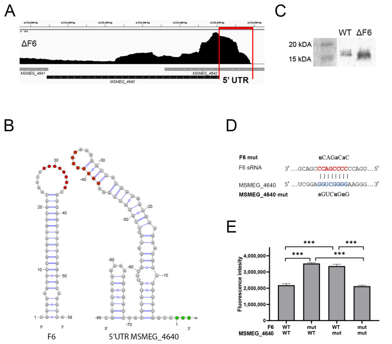
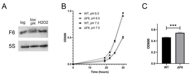
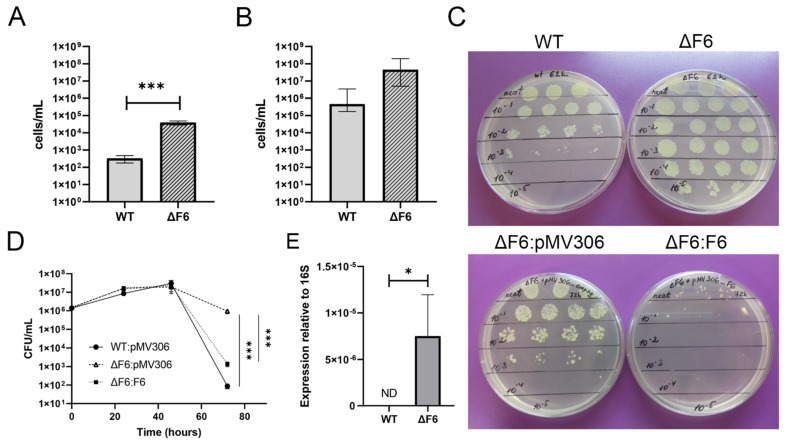
Regulatory small non-coding RNAs play a significant role in bacterial adaptation to changing environmental conditions. Various stresses such as hypoxia and nutrient starvation cause a reduction in the metabolic activity of Mycobacterium smegmatis, leading to entry into dormancy. We investigated the functional role of F6, a small RNA of M. smegmatis, and constructed an F6 deletion strain of M. smegmatis. Using the RNA-seq approach, we demonstrated that gene expression changes that accompany F6 deletion contributed to bacterial resistance against oxidative stress. We also found that F6 directly interacted with 5'-UTR of MSMEG_4640 mRNA encoding RpfE2, a resuscitation-promoting factor, which led to the downregulation of RpfE2 expression. The F6 deletion strain was characterized by the reduced ability to enter into dormancy (non-culturability) in the potassium deficiency model compared to the wild-type strain, indicating that F6 significantly contributes to bacterial adaptation to non-optimal growth conditions.

Keywords: F6; Mycobacterium smegmatis; adaptation to stresses; dormancy; non-culturability; resuscitation promoting factor RpfE2; small non-coding RNA.


Conflict of interest statement

The authors declare no conflict of interest.

Figures

Figure 1
Figure 1
(A) Growth curves of M. smegmatis wt and ΔF6 in Sauton’s medium supplemented with 0.05% (v/v) Tween 80. The data are presented as the mean ± SD of three independent experiments. (B) Light microscopy image of wt and ΔF6 M. smegmatis cells in the early-log phase; magnification ×1250 (left panels) and the image of wt and ΔF6 cultures growing in liquid media (right panels).
Figure 2
Figure 2
RNA-seq of the wt and ΔF6 strains. (A) Volcano plot of differentially expressed genes (DEGs) constructed using Enhanced Volcano R package [24]. Fold changes of gene expression were plotted. Significant DEGs were identified by >four-fold change (log2 FC > 2) and <0.05 FDR, and are shown in red. (B) Schematic representation of the gene clusters upregulated in the ΔF6 strain. (C) Northern blotting analysis of F6 transcription in the wt, ΔF6, and F6 complemented (ΔF6:F6) strains. (D) Validation of DEGs by qRT-PCR. mRNA expression was determined in the wt, ΔF6, ΔF6:pMV306, and ΔF6:F6 cultures in the mid-log phase and normalized to that of 16S rRNA. * p < 0.05, ** p < 0.01, and *** p < 0.001. The data are presented as the mean and SD of three biological replicates for each strain.
Figure 3
Figure 3
F6 of M. smegmatis directly interacts with the 5′-UTR of MSMEG_4640. (A) Inhibition of RpfE2 protein expression by F6. The wt and ΔF6 cultures were analyzed for RpfE2 expression by Western blotting using antibodies against the Rpf conserved domain. The RpfE2 molecular mass is 15.1 kDa according to Mycobrowser data (https://mycobrowser.epfl.ch/genes/MSMEG_4640, accessed on 28 June 2021). (B) The coverage track of the MSMEG_4640 locus in Integrative Genomics Viewer [25]. RNA-seq data of the ΔF6 strain in the mid-log growth phase are shown. The 5′-UTR is marked by vertical red lines. (C) Secondary structures of F6 and the 5′-UTR of MSMEG_4640. The interacted nucleotides are shown as red dots; green dots mark the start codon. (D) Schematic representation of the interaction between F6 and its target MSMEG_4640. The F6 seed region is in red and the complementary 5′-UTR region is in blue. The introduced mutations are shown above and below. (E) The reporter assay illustrating the direct regulation of MSMEG_4640 by F6. The 5′-UTR of MSMEG_4640 was fused to the GFP gene and reciprocal mutations were introduced in the putative interaction sites on F6 and MSMEG_4640-GFP. GFP translation was estimated by fluorescence. The data are presented as the mean ± SD of three biological replicates for each strain; *** p < 0.001.
Figure 4
Figure 4
Effects of oxidative and acidic stresses on the M. smegmatis ΔF6 and wt strains. (A) F6 transcription in the mid-log growth phase and under acidic (low pH) and oxidative (H2O2) stresses was analyzed by Northern blotting. (B) Growth at neutral (pH 7) and acidic (pH 6) conditions. (C) Growth under oxidative stress (0.5 mM H2O2) at eight hours after H2O2 addition. The data are presented as the mean ± SD of three independent experiments; *** p < 0.001.
Figure 5
Figure 5
F6 regulates the transition of M. smegmatis to dormancy under growth-limiting conditions. (A) Colony-forming unit (CFU) concentration for M. smegmatis wt and ΔF6 strains growing in potassium-limiting conditions at the point of minimal culturability (65 h). (B) Reactivation of dormant ‘non-culturable’ M. smegmatis cells in the wt and ΔF6 strains co-cultured with M. luteus at the point of minimal culturability (65 h) in standard Sauton’s medium (most probable numbers, MPN). (C) Culturability of M. smegmatis wt and ΔF6 strains under potassium-limiting conditions. (D) Growth of M. smegmatis wt, ΔF6:pMV306, and ΔF6:F6 strains in potassium-deficient medium. (E) qRT-PCR analysis of MSMEG_4640 expression in wt and ΔF6 cultures growing under potassium limiting conditions (45 h). The data are presented as the mean ± SD of four (A,B,D) or three (E) biological replicates for each strain; * p < 0.05 and *** p < 0.001.

References

    1. Holmqvist E., Wagner E.G.H. Impact of bacterial sRNAs in stress responses. Biochem. Soc. Trans. 2017;45:1203–1212. doi: 10.1042/BST20160363. - DOI - PMC - PubMed
    1. Papenfort K., Vogel J. Regulatory RNA in bacterial pathogens. Cell Host Microbe. 2010;8:116–127. doi: 10.1016/j.chom.2010.06.008. - DOI - PubMed
    1. Waters L.S., Storz G. Regulatory RNAs in bacteria. Cell. 2009;136:615–628. doi: 10.1016/j.cell.2009.01.043. - DOI - PMC - PubMed
    1. Arnvig K.B., Young D.B. Identification of small RNAs in Mycobacterium tuberculosis. Mol. Microbiol. 2009;73:397–408. doi: 10.1111/j.1365-2958.2009.06777.x. - DOI - PMC - PubMed
    1. Gerrick E.R., Barbier T., Chase M.R., Xu R., François J., Lin V.H., Szucs M.J., Rock J.M., Ahmad R., Tjaden B. Small RNA profiling in Mycobacterium tuberculosis identifies MrsI as necessary for an anticipatory iron sparing response. Proc. Natl. Acad. Sci. USA. 2018;115:6464–6469. doi: 10.1073/pnas.1718003115. - DOI - PMC - PubMed

MeSH terms