. 2021 Oct 27;22(21):11633.
doi: 10.3390/ijms222111633.

Targeting Folate Metabolism Is Selectively Cytotoxic to Glioma Stem Cells and Effectively Cooperates with Differentiation Therapy to Eliminate Tumor-Initiating Cells in Glioma Xenografts

Affiliations

Targeting Folate Metabolism Is Selectively Cytotoxic to Glioma Stem Cells and Effectively Cooperates with Differentiation Therapy to Eliminate Tumor-Initiating Cells in Glioma Xenografts

Masashi Okada et al. Int J Mol Sci. .

Abstract

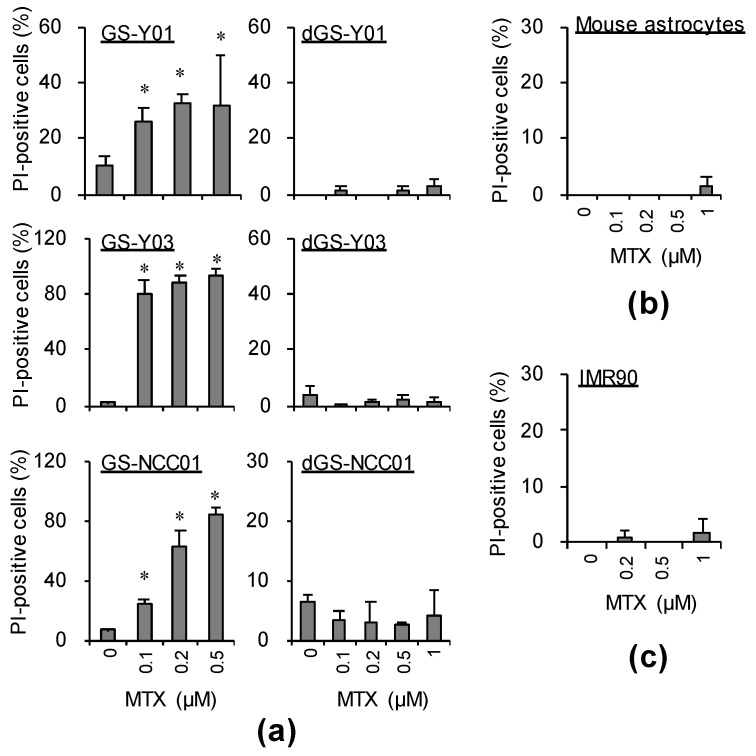
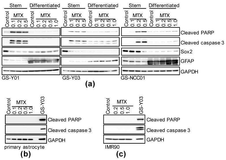
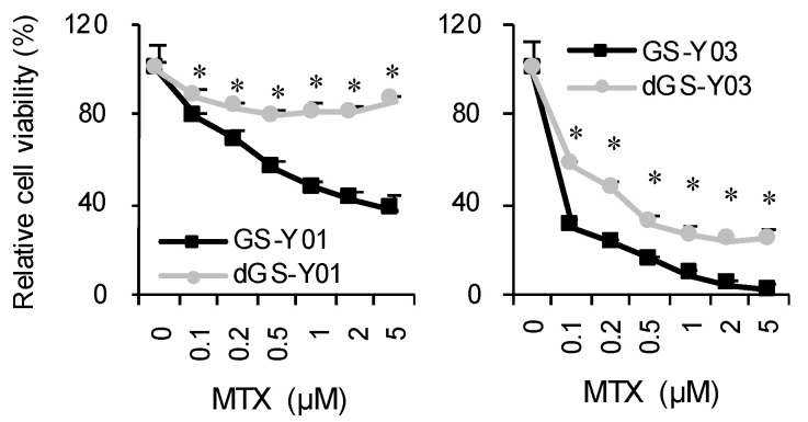
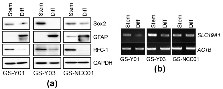
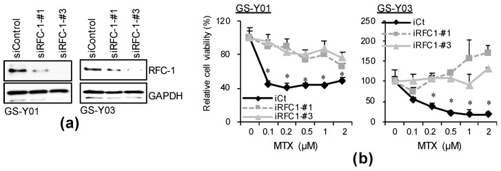
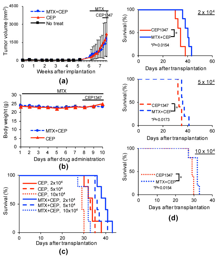
Glioblastoma (GBM) is one of the deadliest of all human cancers. Developing therapies targeting GBM cancer stem cells or glioma stem cells (GSCs), which are deemed responsible for the malignancy of GBM due to their therapy resistance and tumor-initiating capacity, is considered key to improving the dismal prognosis of GBM patients. In this study, we found that folate antagonists, such as methotrexate (MTX) and pemetrexed, are selectively cytotoxic to GSCs, but not to their differentiated counterparts, normal fibroblasts, or neural stem cells in vitro, and that the high sensitivity of GCSs to anti-folates may be due to the increased expression of RFC-1/SLC19A1, the reduced folate carrier that transports MTX into cells, in GSCs. Of note, in an in vivo serial transplantation model, MTX alone failed to exhibit anti-GSC effects but promoted the anti-GSC effects of CEP1347, an inducer of GSC differentiation. This suggests that folate metabolism, which plays an essential role specifically in GSCs, is a promising target of anti-GSC therapy, and that the combination of cytotoxic and differentiation therapies may be a novel and promising approach to effectively eliminate cancer stem cells.

Keywords: JNK; RFC-1; anti-folate; brain tumor initiating cells; glioma stem cell; serial transplantation assay.


Conflict of interest statement

The authors declare no conflict of interest.

Figures

Figure 1
Figure 1
Methotrexate (MTX) induces cell death specifically in glioma stem cells (GSCs), but not in differentiated GSCs. GSCs (GS-Y01, GS-Y03, and GS-NCC01) and differentiated GSCs (dGS-Y01, dGS-Y03, and dGS-NCC01) (a), mouse astrocytes (b), and IMR90 cells (c) treated with the indicated concentrations of methotrexate (MTX) or control solvent treatment for 3 days were analyzed by propidium iodide incorporation assay. Similar results were obtained from two independent biological replicates. Data are presented as means + standard deviation. * p < 0.05 vs. control-treated cells by the Mann–Whitney U test.
Figure 2
Figure 2
MTX induces caspase-activated cell death in GSCs, but not in differentiated GSCs. GSCs (GS-Y01, GS-Y03, GS-NCC01; Stem or Differentiated, (a)), mouse primary astrocytes (b), and IMR90 (c) treated with the indicated concentrations (μM) of MTX for 3 days or control-treated were analyzed by immunoblotting for the indicated proteins. “GS-Y03” in (b,c) denotes GS-Y03 cells treated with 0.2 μM MTX for 3 days. Representative images of two independent biological replicates.
Figure 3
Figure 3
Growth-inhibitory effects of MTX on GSCs and differentiated GSCs. GSCs (GS-Y01, GS-Y03) and differentiated GSCs (dGS-Y01, dGS-Y03) treated with MTX at the indicated concentrations for 3 days were subjected to the WST-8 assay. Their viability relative to control was measured. Values in the graphs represent means + standard deviation from triplicate samples of a representative experiment. Similar results were obtained from two independent biological replicates. * p < 0.05 vs. each concentration MTX-treated GSCs by Student’s t-test.
Figure 4
Figure 4
The expression of RFC-1/SLC19A1 in GSCs is reduced by differentiation. GSCs (GS-Y01, GS-Y03, and GS-NCC01; Stem) and differentiated GSCs (Diff) were analyzed by immunoblotting (a) or RT-PCR (b) for the indicated proteins or mRNAs. Representative images of two biological replicates are shown.
Figure 5
Figure 5
Selective toxicity of MTX to GSCs is dependent on RFC-1 expression. (a) GS-Y01 and GS-Y03 cells (2–5 × 10⁵) were transiently transfected with an siRNA against RFC-1 (siRFC-1) or with control siRNA (siControl) and cultured for 4 days. The cells were subjected to immunoblotting analysis for RFC-1 and GAPDH. (b) The siRNA-transfected cells were treated with MTX at the indicated concentrations for 3 days and then subjected to WST-8 analysis. * p < 0.05 vs. siControl-transfected cells treated with each concentration of MTX by Dunnett’s test.
Figure 6
Figure 6
Systemic administration of MTX cooperates with CEP1347 to inhibit tumor-initiating cells in tumors in tumor-bearing mice. (a,b) Mice implanted subcutaneously with GS-Y03 (1 × 10⁶ cells) were randomized into 2 treatment groups (3 mice per group) 6 weeks after implantation, when the average primary tumor volume reached approximately 300 mm3, and received a daily intraperitoneal injection of CEP1347 (CEP; 1.5 mg/kg/day) for 3 consecutive days, as indicated schematically in (a,b). Mice in the MTX + CEP group received an additional daily intraperitoneal injection of MTX (50 mg/kg/day) followed by intraperitoneal leucovorin (50 mg/kg/day, 4 h after MTX) for 10 consecutive days, which started at the time of randomization and 1 week before the administration of CEP1347. In the combination (MTX + CEP) group, MTX was administered 6 h before CEP1347. One day after the final drug treatment, the subcutaneous tumors were excised and dissociated. Then, the serial dilutions of the dissociated tumor cells were transplanted intracranially into new mice. The volume of the primary tumors treated with CEP1347 or MTX + CEP1347 (MTX + CEP) was assessed at the indicated time points and is presented in the graph as the mean ± SD (a), and body weight was monitored at the indicated time points (b). Kaplan–Meier survival curves of the mice (n = 5 for each group) are shown (c). For clarity, the survival curves of mice transplanted with 2 × 10⁴ (upper), 5 × 10⁴ (middle), or 10 × 104 (lower) cells are shown separately in (d). * p < 0.05 vs. CEP1347-treated mice by the log-rank test.

Similar articles

Cited by

References

    1. Matsuda K., Sato A., Okada M., Shibuya K., Seino S., Suzuki K., Watanabe E., Narita Y., Shibui S., Kayama T., et al. Targeting JNK for therapeutic depletion of stem-like glioblastoma cells. Sci. Rep. 2012;2:516. doi: 10.1038/srep00516. - DOI - PMC - PubMed
    1. Okada M., Kuramoto K., Takeda H., Watarai H., Sakaki H., Seino S., Seino M., Suzuki S., Kitanaka C. The novel JNK inhibitor AS602801 inhibits cancer stem cells in vitro and in vivo. Oncotarget. 2016;7:27021–27032. doi: 10.18632/oncotarget.8395. - DOI - PMC - PubMed
    1. Okada M., Takeda H., Sakaki H., Kuramoto K., Suzuki S., Sanomachi T., Togashi K., Seino S., Kitanaka C. Repositioning CEP-1347, a chemical agent originally developed for the treatment of Parkinson’s disease, as an anti-cancer stem cell drug. Oncotarget. 2017;8:94872–94882. doi: 10.18632/oncotarget.22033. - DOI - PMC - PubMed
    1. Sato A., Sunayama J., Okada M., Watanabe E., Seino S., Shibuya K., Suzuki K., Narita Y., Shibui S., Kayama T., et al. Glioma-initiating cell elimination by metformin activation of FOXO3 via AMPK. Stem Cells Transl. Med. 2012;1:811–824. doi: 10.5966/sctm.2012-0058. - DOI - PMC - PubMed
    1. Shibuya K., Okada M., Suzuki S., Seino M., Seino S., Takeda H., Kitanaka C. Targeting the facilitative glucose transporter GLUT1 inhibits the self-renewal and tumor-initiating capacity of cancer stem cells. Oncotarget. 2015;6:651–661. doi: 10.18632/oncotarget.2892. - DOI - PMC - PubMed

MeSH terms