Review
. 2021 Oct 30;22(21):11799.
doi: 10.3390/ijms222111799.

Transient Fluorescence Labeling: Low Affinity-High Benefits

Affiliations
Review

Transient Fluorescence Labeling: Low Affinity-High Benefits

Maxim M Perfilov et al. Int J Mol Sci. .

Abstract

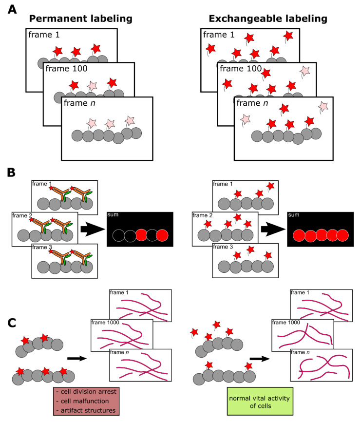
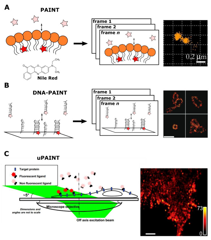
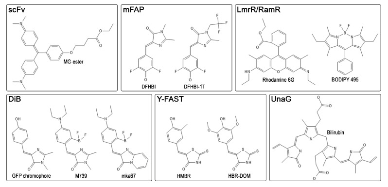
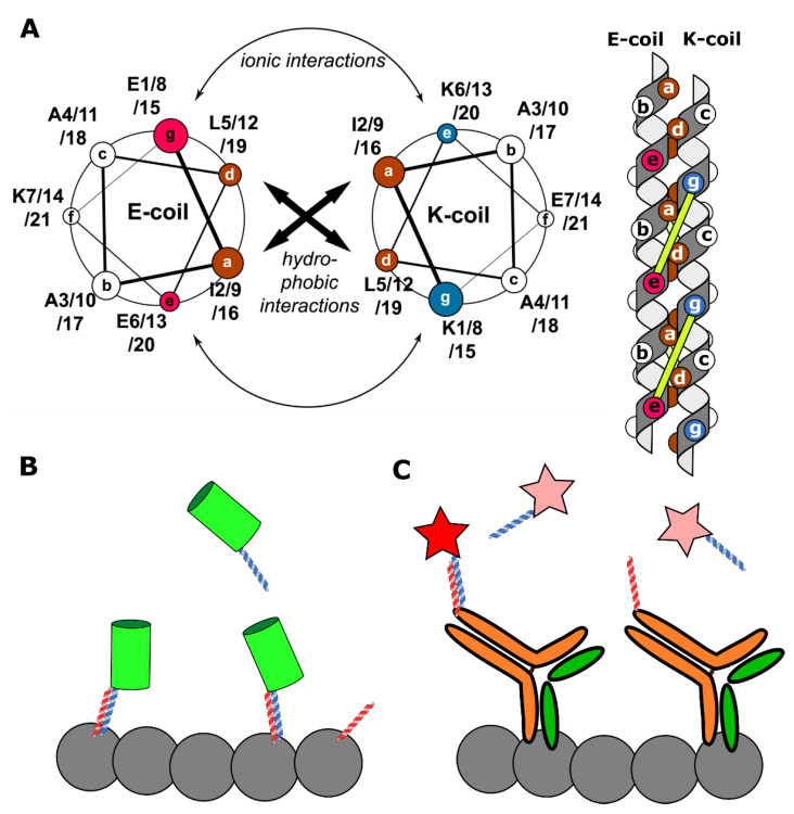
Fluorescent labeling is an established method for visualizing cellular structures and dynamics. The fundamental diffraction limit in image resolution was recently bypassed with the development of super-resolution microscopy. Notably, both localization microscopy and stimulated emission depletion (STED) microscopy impose tight restrictions on the physico-chemical properties of labels. One of them-the requirement for high photostability-can be satisfied by transiently interacting labels: a constant supply of transient labels from a medium replenishes the loss in the signal caused by photobleaching. Moreover, exchangeable tags are less likely to hinder the intrinsic dynamics and cellular functions of labeled molecules. Low-affinity labels may be used both for fixed and living cells in a range of nanoscopy modalities. Nevertheless, the design of optimal labeling and imaging protocols with these novel tags remains tricky. In this review, we highlight the pros and cons of a wide variety of transiently interacting labels. We further discuss the state of the art and future perspectives of low-affinity labeling methods.

Keywords: PAINT; exchangeable labels; fluorescent labeling; super-resolution microscopy.


Conflict of interest statement

The authors declare no conflict of interest.

Figures

Figure 1
Figure 1
Comparison of permanent (high-affinity or covalent) and exchangeable (transient) labeling. (A) Permanent labels are constantly exposed to light irradiation and inevitably photobleached. Therefore, the fluorescent signal deteriorates, making prolonged imaging more complicated. In contrast, the continuous exchange of low-affinity labels with undamaged ones from cytosol or medium increases the apparent photostability of labeling. Red stars denote fluorescent labels, pink stars denote photobleached labels. (B) High-affinity labels already bound to target structures, due to their large size, can sterically interfere with the binding of other label molecules. Alternatively, frame-by-frame accumulation of low-affinity labels’ positions followed by frames merging increases effective labeling density. (C) Bulky labels, continuously bound to target structures, may affect the dynamics and functioning of the latter. In addition, some labels, such as fluorescently labeled taxol or fluorescent proteins could drastically disturb cell activity. However, in the case of low-affinity labeling, target molecules remain untagged most of the time and therefore their functioning is less hindered.
Figure 2
Figure 2
A schematic illustration of PAINT methods. (A) An original PAINT approach. Left: an environment-sensitive probe (Nile Red) fluorescently bursts upon reversible interaction with the lipid layer. Center: accumulation of fluorescent bursts during n frames registration. Right: the reconstructed image of vesicles, imaged with PAINT approach using Nile Red probe. Reprinted from Sharonov et al. [26] (copyright (2006) National Academy of Sciences). (B) A DNA-PAINT approach. Left: in the DNA-PAINT labeling system target and dye molecules are conjugated to complementary DNA strands. Transient interaction between strands temporarily co-localizes fluorescent probes with a target structure. This binding event is detected as a burst of fluorescence. Center: accumulation of fluorescent bursts during n frames registration. Right: reconstructed images of long rectangular DNA-origami oligomers labeled with DNA-PAINT. Scale bar 500 nm. Reprinted with permission from Jungmann et al. [30]. Copyright (2010) American Chemical Society. (C) A uPAINT approach. Left: fluorescent probes bind to target sites in the cell. A low-angle excitation laser beam (angle of about 5°) illuminates ≈2 μm thick cross section, thereby excites predominantly only bound labels. Right: Super-resolved image of the transmembrane protein TM-6His labeled with trisNTA-AT647N obtained by uPAINT. Scale bar 1 μm. Reprinted from Giannone et al. [31], copyright (2010), with permission from Elsevier.
Figure 3
Figure 3
Common representatives of ligands for known FAP-based labeling systems. MG-ester—malachite green ester, DFHBI—difluoro-4-hydroxybenzylidene imidazolinone, DFHBI-1T—3,5-difluoro-4-hydroxybenzylidene-2,2,2-trifluoroethyl imidazolinone, BODIPY—4,4-difluoro-4-bora-3a,4a-diaza-s-indacene, HMBR—4-hydroxy-3-methylbenzylidene rhodanine, HBR-DOM—4-hydroxy-3,5-dimethoxybenzylidene rhodanine.
Figure 4
Figure 4
A schematic representation of K/E-coils labeling systems. (A) Left: a helical wheel diagram, demonstrating the amino acid residues in interacting helices (reprinted by permission from Springer Nature Customer Service Centre Gmbh: Springer Nature Cellular and Molecular Life Sciences, Perfilov et al. [21], Copyright Clearance Center (2020)). Heptad positions are labeled with ag. Brown positions show hydrophobic amino acid residues, purple—positively charged residues, and blue—negatively charged residues. Right: a schematic side view representation of a coiled-coil complex. Circles on helices show amino acid residues from the helical wheel diagram. The complex is stabilized both by hydrophobic interactions between a and d positions and ionic interactions between residues i (position g) of one helix and residues i + 5 of the other helix (position e). Ionic interactions are represented by yellow lines. (B) The KECs approach. Target proteins (grey circles) are tagged by E-coils (red helices). K-coils (blue helices) are bound to fluorescent proteins (green cylinder). Reversible interaction between K- and E-coils makes possible target proteins visualization. In contrast, in the peptide-PAINT approach (C) E-coils are conjugated to target-specific antibodies and K-coils are Cy3B labeled. Similar to the DNA-PAINT, labels are only visible (red star) when a coiled-coil complex is formed, while unbound labels are undetectable (pink stars) [108].

References

    1. Sahl S.J., Hell S.W., Jakobs S. Fluorescence nanoscopy in cell biology. Nat. Rev. Mol. Cell Biol. 2017;18:685–701. doi: 10.1038/nrm.2017.71. - DOI - PubMed
    1. Tsien R.Y. The green fluorescent protein. Annu. Rev. Biochem. 1998;67:509–544. doi: 10.1146/annurev.biochem.67.1.509. - DOI - PubMed
    1. Mishin A.S., Belousov V.V., Solntsev K.M., Lukyanov K.A. Novel uses of fluorescent proteins. Curr. Opin. Chem. Biol. 2015;27:1–9. doi: 10.1016/j.cbpa.2015.05.002. - DOI - PubMed
    1. Hoelzel C.A., Zhang X. Visualizing and manipulating biological processes by using HaloTag and SNAP-Tag technologies. ChemBioChem. 2020;21:1935–1946. doi: 10.1002/cbic.202000037. - DOI - PMC - PubMed
    1. Xu J., Ma H., Liu Y. Stochastic Optical Reconstruction Microscopy (STORM) Curr. Protoc. Cytom. 2017;81:12.46.1–12.46.27. doi: 10.1002/cpcy.23. - DOI - PMC - PubMed

Substances

LinkOut - more resources