Review
. 2021 Oct 30;22(21):11810.
doi: 10.3390/ijms222111810.

Deacetylation of Transcription Factors in Carcinogenesis

Affiliations
Review

Deacetylation of Transcription Factors in Carcinogenesis

Marta Halasa et al. Int J Mol Sci. .

Abstract

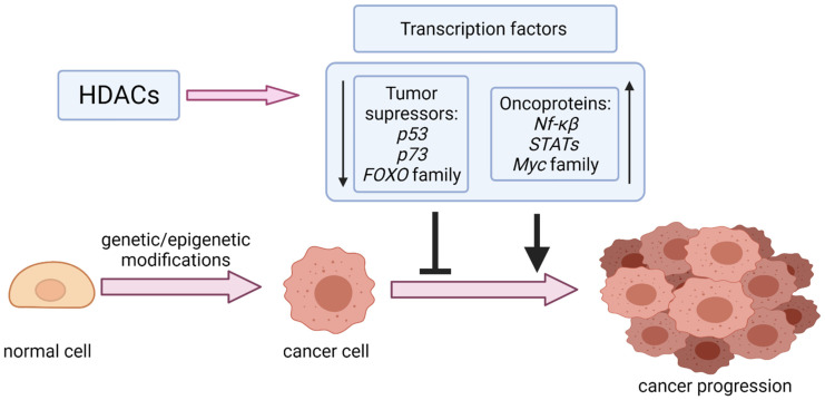
Reversible Nε-lysine acetylation/deacetylation is one of the most common post-translational modifications (PTM) of histones and non-histone proteins that is regulated by histone acetyltransferases (HATs) and histone deacetylases (HDACs). This epigenetic process is highly involved in carcinogenesis, affecting histone and non-histone proteins' properties and their biological functions. Some of the transcription factors, including tumor suppressors and oncoproteins, undergo this modification altering different cell signaling pathways. HDACs deacetylate their targets, which leads to either the upregulation or downregulation of proteins involved in the regulation of cell cycle and apoptosis, ultimately influencing tumor growth, invasion, and drug resistance. Therefore, epigenetic modifications are of great clinical importance and may constitute a new therapeutic target in cancer treatment. This review is aimed to present the significance of HDACs in carcinogenesis through their influence on functions of transcription factors, and therefore regulation of different signaling pathways, cancer progression, and metastasis.

Keywords: HDAC; histone deacetylase inhibitors (HDIs); transcription factors.


Conflict of interest statement

The authors declare no conflict of interest.

Figures

Figure 1
Figure 1
Modulation of transcription factors (tumor suppressors and oncoproteins) by HDACs in cancer progression.
Figure 2
Figure 2
The biological effects of the p53-Ac and p73-Ac deacetylation catalyzed by SIRTs and HDACs. ↑—upregulation, ↓—downregulation (A) BMRS1 disrupts the interaction between SIRT1 and DBC1, decreasing the p53-Ac level. (B) Phosphorylated DBC1 binds to SIRT1 that dissociates the SIRT1–p53 complex and simulates p53 acetylation leading to apoptosis. (C) BRG1 binds to SIRT1, which results in increased p53-Ac deacetylation and inhibits p53/p21-mediated cell senescence. (D) Downregulation of SIRT1 expression by specific inhibitors is associated with the upregulation of p53-Ac, which leads to the upregulation of proteins involved in apoptosis. (E) Repression of miR-34a/SIRT1/p53 feedback loop by HNF1A-AS1 leads to a decrease in p53-Ac and activation of the Wtn signaling pathway, which, in turn, enhances cancer progression. (F) SIRT1 is regulated by several miRNAs, leading to a change of the p53-Ac level and modulation of apoptosis. (G) The influence of SIRT3 on p53Ac deacetylation and p53 degradation by the proteasome. (H) SIRT6 deacetylates p53-Ac, which is ubiquitinated and degraded by the proteasome. (I) SIRT7 deacetylates p53-Ac, which leads to downregulation of Noxa and ultimately the inhibition of cell apoptosis. (J) SIRT1, SIRT2, and HDAC1 can deacetylate p73-Ac, leading to a decrease in apoptosis and increased cell proliferation and tumorigenicity.BMRS1—breast cancer metastasis suppressor 1; DBC1—breast cancer 1; BRG1—brahma-related gene-1; HNF1A-AS1—HNF1A-antisense 1 RNA1.
Figure 3
Figure 3
The interplay between SIRTs, HDACs, and FOXOs and the biological results of these interactions. ↑—upregulation, ↓—downregulation (A) Downregulation of SIRT1 and SIRT7 by LPS treatment increases FOXO3a acetylation and phosphorylation, which induces apoptosis. (B) Akt inhibitors interrupt phosphorylated FOXO3a–SIRT6 interaction, leading to FOXO3a acetylation and BRD4 binding, promoting upregulation of CDK6, cancer progression, and drug resistance. (C) FOXO3a is deacetylated by HDAC3, which leads to Dicer downregulation and enhances metastasis. (D) SIRT1 deacetylates FOXO1, leading to MRP2 upregulation and enhanced drug resistance. (E) Downregulation of SIRT1 upregulates FOXO1, leading to increased sensitivity to progesterone therapy, and modulates expression of genes involved in the regulation of cancer growth, angiogenesis, cell survival, EMT, cell proliferation, and cancer invasion.LPS—lipopolysaccharide; BRD4—bromodomain-containing protein 4; CDK6—cyclin dependent kinase 6; MRP2—multidrug resistance protein 2; EMT—epithelial-mesenchymal transition.
Figure 4
Figure 4
The influence of HDACs and SIRTs on NF-κB and biological results of these modifications. ↑—upregulation, ↓—downregulation, ¬ inhibition, (A) SIRT1 deacetylates MYST1-Ac, which interacts with p65. The MYST1–p65 complex binds SIRT1 or AR, thus modulating the intensity of cell proliferation and apoptosis. (B) SIRT2 deacetylates p65-Ac, leading to downregulation of miR-21 expression and inhibition of cancer growth. (C) Downregulation of SIRT7 leads to a decrease in N.F.- κB, Bcl-xl, Bcl-2, and Mcl-1 expressions, and the upregulation of caspase-3 and Bad and BAX proteins induces apoptosis and inhibits the growth and invasiveness of cancer cells. (D) HDAC6 deacetylates p65-Ac, preventing p65 from binding to the MMP2 promoter region and ultimately decreasing cancer invasiveness.AR—androgen receptor; N.F.- κB—nuclear factor kappa-light-chain-enhancer of activated B cells; Mcl-1—myeloid cell leukaemia-1; MMP2- matrix metalloproteinase-2.
Figure 5
Figure 5
The influence of SIRTs and HDACs on STAT deacetylation and biological results of these modifications. ↑—upregulation, ↓—downregulation. (A) HDAC1 and HDAC4 deacetylate STAT3-Ac leading to decrease in STAT3 transcriptional activity; (B) SIRT1 represses FGB leading to inhibition of cell proliferation; (C) Downregulation of SIRT1 results in upregulation of STAT3-Ac and MMP-13 what enhances cell proliferation.STAT—signal transducers and activators of transcription; FGB—fibrinogen beta chain; MMP13-matrix metalloproteinase 13.
Figure 6
Figure 6
The interplay between SIRT1 and Myc proteins. (A) SIRT1 deacetylates C-Myc and thus enables the formation of C-Myc/Max heterodimers, resulting in C-Myc transactivation, followed by an increased expression of its target genes and thus stimulation of cell growth and proliferation. Simultaneously, C-Myc stimulates the activity of SIRT1 by inducing the expression of the NAMPT gene encoding the enzyme responsible for the production of the SIRT1 cofactor NAD+. (B) The use of the NAM leads to the inhibition of SIRT1 activity and thus inhibits C-Myc protein deacetylation, decreases the expression of target genes, and results in cell cycle arrest. NAMPT—nicotinamide-phosphoribosyltransferase; NAD+—nicotinamide adenine dinucleotide; hTERT—human telomerase reverse transcriptase; NAM—nicotinamide.

References

    1. Sung H., Ferlay J., Siegel R.L., Laversanne M., Soerjomataram I., Jemal A., Bray F. Global cancer statistics 2020: GLOBOCAN estimates of incidence and mortality worldwide for 36 cancers in 185 countries. CA Cancer J. Clin. 2021;71:209–249. doi: 10.3322/caac.21660. - DOI - PubMed
    1. Ahmad F.B., Anderson R.N. The Leading Causes of Death in the US for 2020. JAMA. 2021;325:1829–1830. doi: 10.1001/jama.2021.5469. - DOI - PMC - PubMed
    1. (US) NI of H, Study BSC Understanding Cancer. [(accessed on 3 September 2021)];2007 Available online: https://www.ncbi.nlm.nih.gov/books/NBK20362/
    1. Jin J., Wu X., Yin J., Li M., Shen J., Li J., Zhao Y., Zhao Q., Wu J., Wen Q. Identification of Genetic Mutations in Cancer: Challenge and Opportunity in the New Era of Targeted Therapy. Front. Oncol. 2019;9:263. doi: 10.3389/fonc.2019.00263. - DOI - PMC - PubMed
    1. Sharma S., Kelly T.K., Jones P.A. Epigenetics in cancer. Carcinogenesis. 2009;31:27–36. doi: 10.1093/carcin/bgp220. - DOI - PMC - PubMed