. 2021 Nov 2;22(21):11911.
doi: 10.3390/ijms222111911.

Fibroblast Differentiation and Matrix Remodeling Impaired under Simulated Microgravity in 3D Cell Culture Model

Affiliations

Fibroblast Differentiation and Matrix Remodeling Impaired under Simulated Microgravity in 3D Cell Culture Model

Jiranuwat Sapudom et al. Int J Mol Sci. .

Abstract

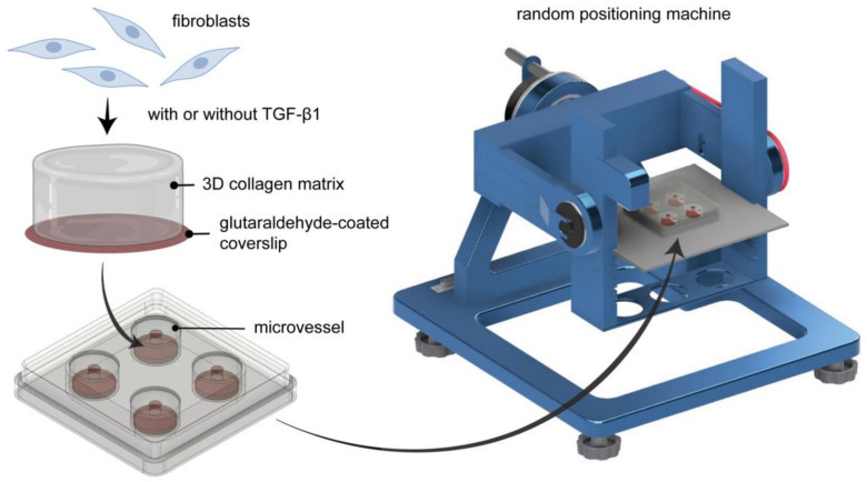
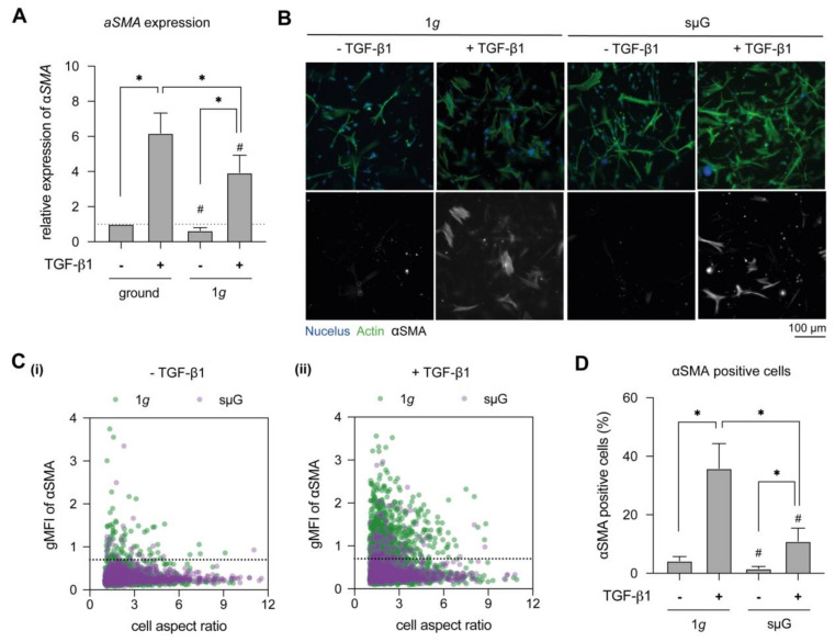
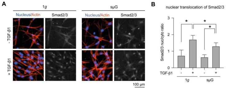
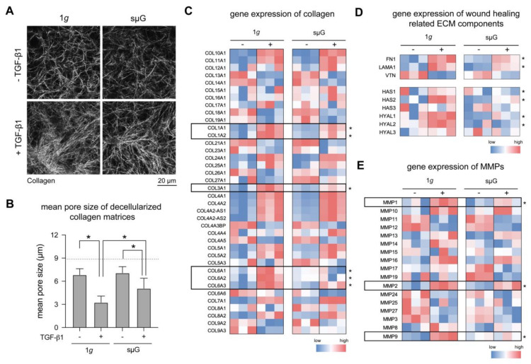
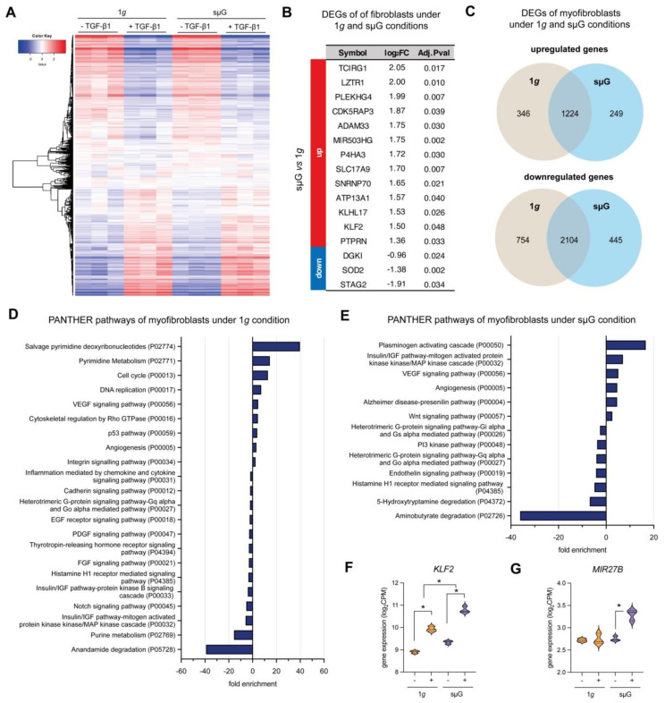
Exposure to microgravity affects astronauts' health in adverse ways. However, less is known about the extent to which fibroblast differentiation during the wound healing process is affected by the lack of gravity. One of the key steps of this process is the differentiation of fibroblasts into myofibroblasts, which contribute functionally through extracellular matrix production and remodeling. In this work, we utilized collagen-based three-dimensional (3D) matrices to mimic interstitial tissue and studied fibroblast differentiation under simulated microgravity (sµG). Our results demonstrated that alpha-smooth muscle actin (αSMA) expression and translocation of Smad2/3 into the cell nucleus were reduced upon exposure to sµG compared to the 1g control, which suggests the impairment of fibroblast differentiation under sµG. Moreover, matrix remodeling and production were decreased under sµG, which is in line with the impaired fibroblast differentiation. We further investigated changes on a transcriptomic level using RNA sequencing. The results demonstrated that sµG has less effect on fibroblast transcriptomes, while sµG triggers changes in the transcriptome of myofibroblasts. Several genes and biological pathways found through transcriptome analysis have previously been reported to impair fibroblast differentiation. Overall, our data indicated that fibroblast differentiation, as well as matrix production and remodeling, are impaired in 3D culture under sµG conditions.

Keywords: 3D cell culture; fibroblast differentiation; matrix remodeling; microgravity; tissue repair.


Conflict of interest statement

The authors declare no conflict of interest.

Figures

Figure 1
Figure 1
Schematic illustration of experimental setup. Fibroblasts were cultured in 3D collagen matrices and placed inside engineered biocompatible microvessels before being cultured either on 1g or on the random positioning machine (RPM). RPM is placed in a conventional cell culture incubator.
Figure 2
Figure 2
Myofibroblast differentiation in dependence of TGF-β1 stimulation under 1g and sµG. Fibroblasts were cultured for 3 days in the presence and absence of TGF-β1 under 1g and sμG conditions. (A) Cells were analyzed regarding αSMA expression using RT-qPCR. Gene expression analysis was performed in four replicates. (B) Representative image of fibroblasts stained with DAPI (blue), Phalloidin (green) and αSMA (grey) antibodies. (C) Quantitative analysis of geometric mean of fluorescence intensity (gMFI) of αSMA and cell aspect ratio using a custom-made image analysis toolbox. The dashed line in C(i) and C(ii) represents the cut-off value, below which are the majority of low αSMA expressing cells without TGF-β1 under 1g condition. (D) Percentage of αSMA positive cells. The image analysis was performed at least in triplicate with four positions per sample. Data are shown as mean +/− SD. * indicates significant p ≤ 0.005. The character # represents the significance level of p ≤ 0.05 when compared to 1g at similar condition.
Figure 3
Figure 3
Nuclear translocation of Smad2/3 in dependence of TGF-β1 stimulation under 1g and sµG. (A) Representative image of cells stained with Hoechst-33342 (blue), Phalloidin (red) and Smad2/3 (grey) antibodies. (B) The quantitative image analysis was performed at least in triplicate with four positions per sample. Data are shown as mean +/− SD. * indicates significant p ≤ 0.005.
Figure 4
Figure 4
Matrix remodeling and ECM gene expression under 1g and sµG conditions. (A) Representative images of decellularized collagen matrices. Matrices were analyzed regarding (B) pore size. The image analysis was performed at least in triplicate with four positions per sample. Data are shown as mean +/− SD. * indicates significant p ≤ 0.05. Gene expression using RNA-Seq data of (C) collagens, (D) wound healing related ECM factors, and (E) matrix metalloproteinases (MMPs) in both 1g and sμG conditions. Data are shown as a heatmap using colors on a scale from red (high expression) to blue (low expression). Statistical significance test was performed for TGF-β1-treated samples between 1g and sµG conditions for Figure 4C–E, which is indicated by * for p-value ≤ 0.05. The experiment was performed in three replicates.
Figure 5
Figure 5
RNA-Sequencing analysis of fibroblast and myofibroblast under 1g and sµG conditions. (A) Heat map of overall gene expression levels in fibroblasts and myofibroblasts under 1g and under sµG. DEGs were analyzed using DESeq2 with FDR cutoff ≤ 0.05 and FC ≥ 2.0 using DESeq2 (B) Table of differentially expressed genes (DEGs) with log2 fold change (log2FC) and adjusted P-value of fibroblasts cultured under 1g and under sµG. Up- and downregulated genes were compared between sµG and 1g conditions. (C) Venn diagram of up- and down-regulated DEGs of myofibroblast cultured under 1g and under sµG. The DEGs were analyzed for enriched biological pathways using statistical overrepresentation tests using the PANTHER pathway for (D) 1g and (E) sµG conditions. Gene expression of specific DEGs which has been reported to be involved in inhibition of fibroblast differentiation, namely (F) KLF2 and (G) MIR27B. Data are shown as a violin plot; * significance level of p < 0.05. The experiment was performed with three replicates.

References

    1. ElGindi M., Sapudom J., Ibrahim I.H., Al-Sayegh M., Chen W., Garcia-Sabaté A., Teo J.C.M. May the Force Be with You (Or Not): The Immune System under Microgravity. Cells. 2021;10:1941. doi: 10.3390/cells10081941. - DOI - PMC - PubMed
    1. Roy-O’Reilly M., Mulavara A., Williams T. A review of alterations to the brain during spaceflight and the potential relevance to crew in long-duration space exploration. NPJ Microgravity. 2021;7:5. doi: 10.1038/s41526-021-00133-z. - DOI - PMC - PubMed
    1. Stavnichuk M., Mikolajewicz N., Corlett T., Morris M., Komarova S.V. A systematic review and meta-analysis of bone loss in space travelers. NPJ Microgravity. 2020;6:13. doi: 10.1038/s41526-020-0103-2. - DOI - PMC - PubMed
    1. Radek K.A., Baer L.A., Eckhardt J., DiPietro L.A., Wade C.E. Mechanical unloading impairs keratinocyte migration and angiogenesis during cutaneous wound healing. J. Appl. Physiol. 2008;104:1295–1303. doi: 10.1152/japplphysiol.00977.2007. - DOI - PubMed
    1. Monici M., Cialdai F., Romano G., Fusi F., Egli M., Pezzatini S., Morbidelli L. An in Vitro Study on Tissue Repair: Impact of Unloading on Cells Involved in the Remodelling Phase. Microgravity Sci. Technol. 2011;23:391–401. doi: 10.1007/s12217-011-9259-4. - DOI

LinkOut - more resources