Comparative Study
. 2021 Nov 4;22(21):11961.
doi: 10.3390/ijms222111961.

The Presence of Yin-Yang Effects in the Migration Pattern of Staurosporine-Treated Single versus Collective Breast Carcinoma Cells

Affiliations
Comparative Study

The Presence of Yin-Yang Effects in the Migration Pattern of Staurosporine-Treated Single versus Collective Breast Carcinoma Cells

Frank A H Meyer et al. Int J Mol Sci. .

Abstract

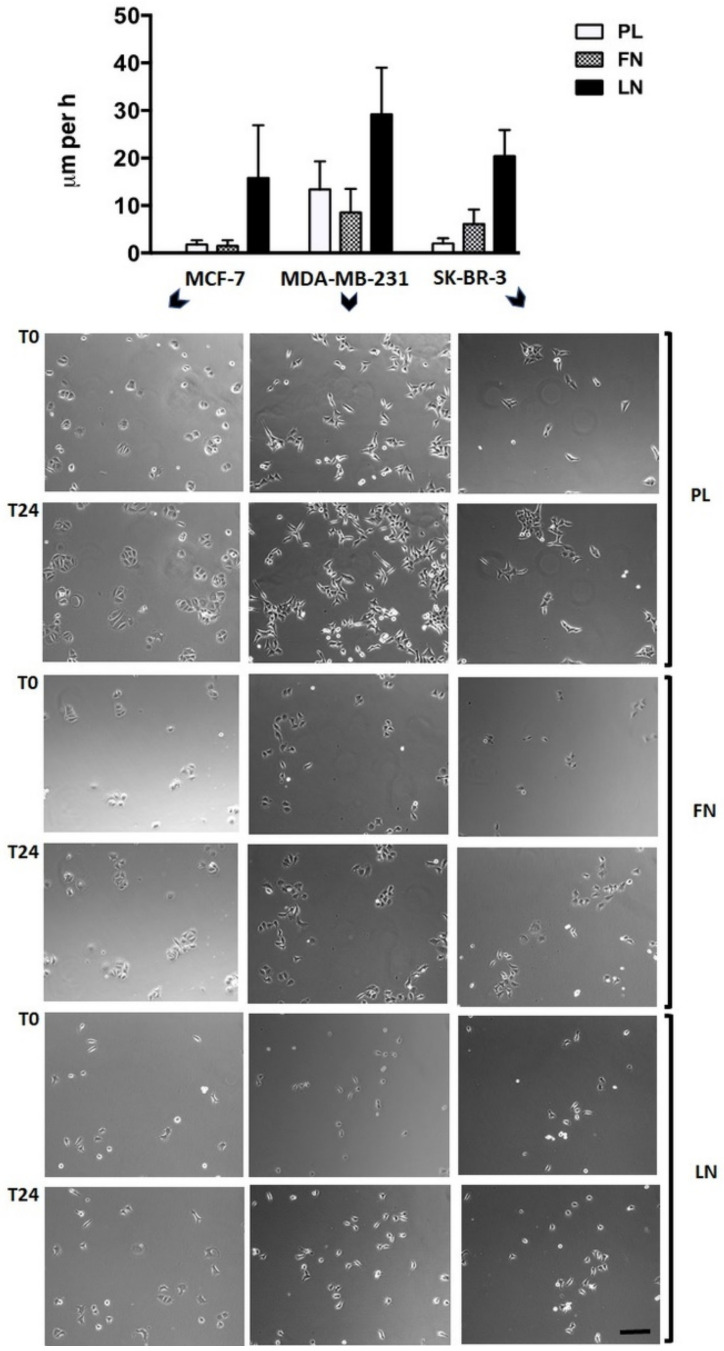
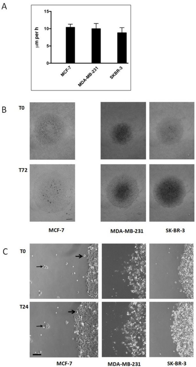
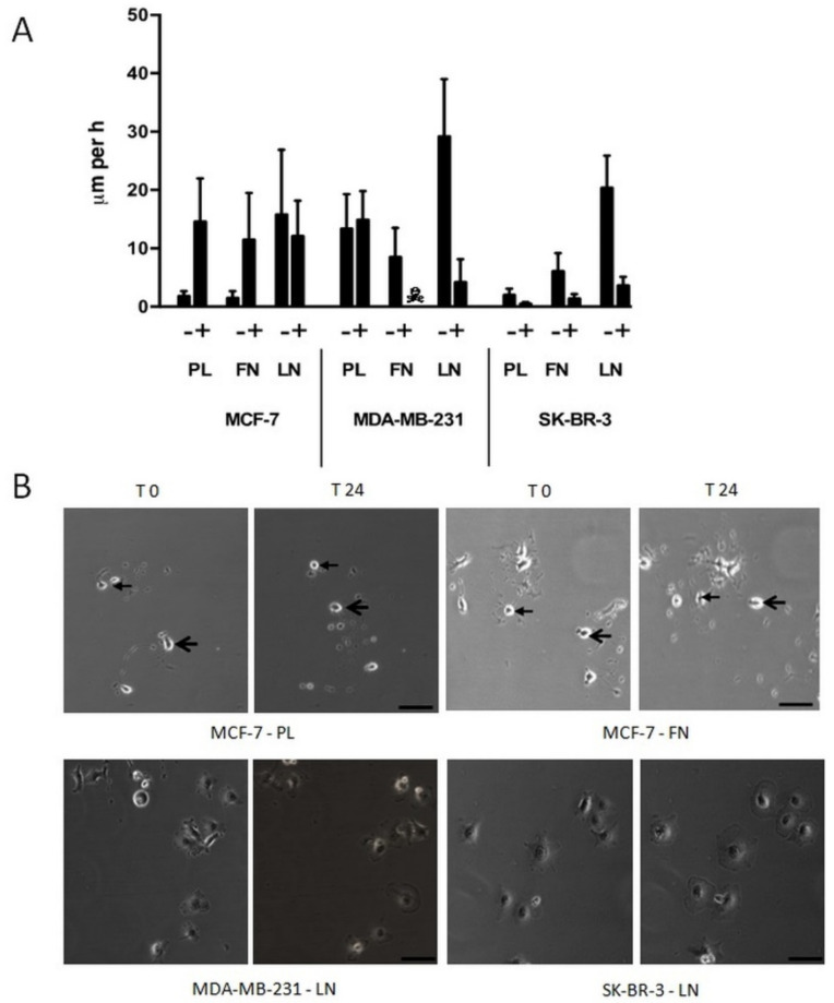
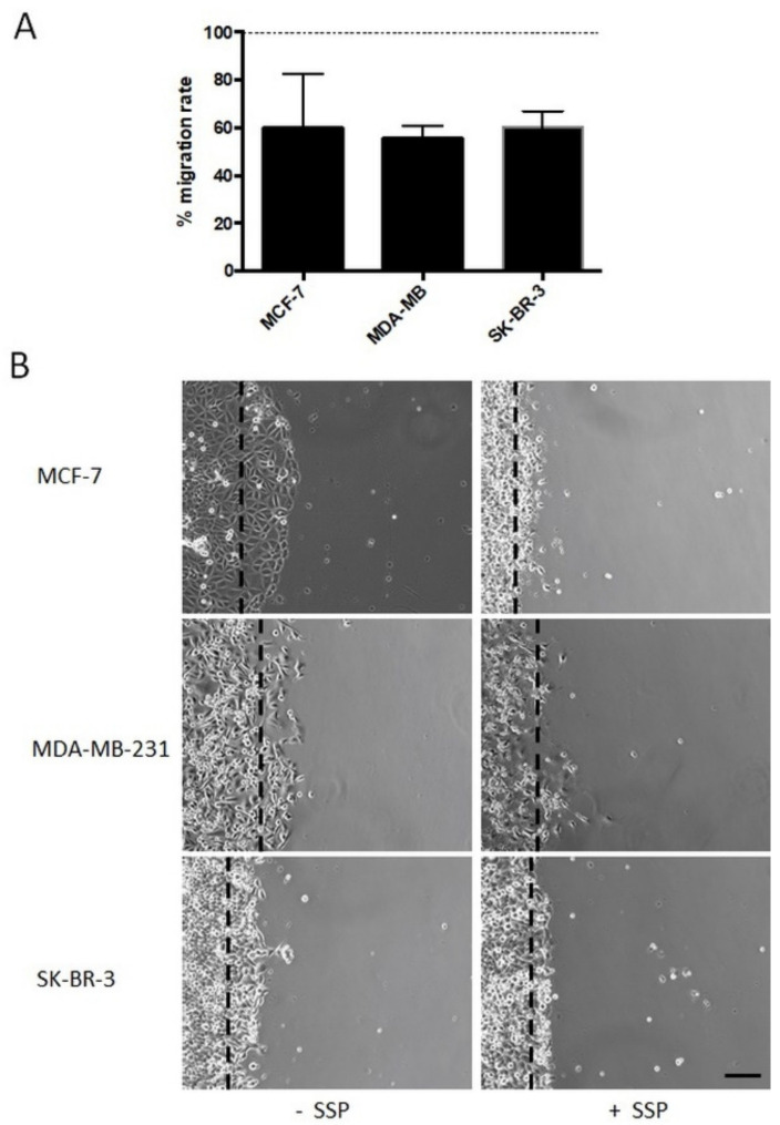
Background: Staurosporine-dependent single and collective cell migration patterns of breast carcinoma cells MDA-MB-231, MCF-7, and SK-BR-3 were analysed to characterise the presence of drug-dependent migration promoting and inhibiting yin-yang effects.

Methods: Migration patterns of various breast cancer cells after staurosporine treatment were investigated using Western blot, cell toxicity assays, single and collective cell migration assays, and video time-lapse. Statistical analyses were performed with Kruskal-Wallis and Fligner-Killeen tests.

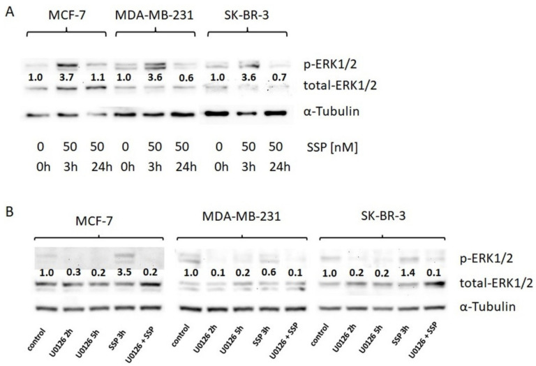
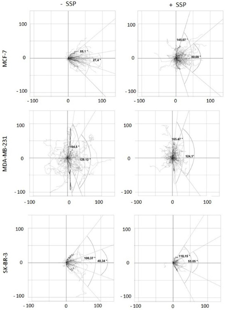
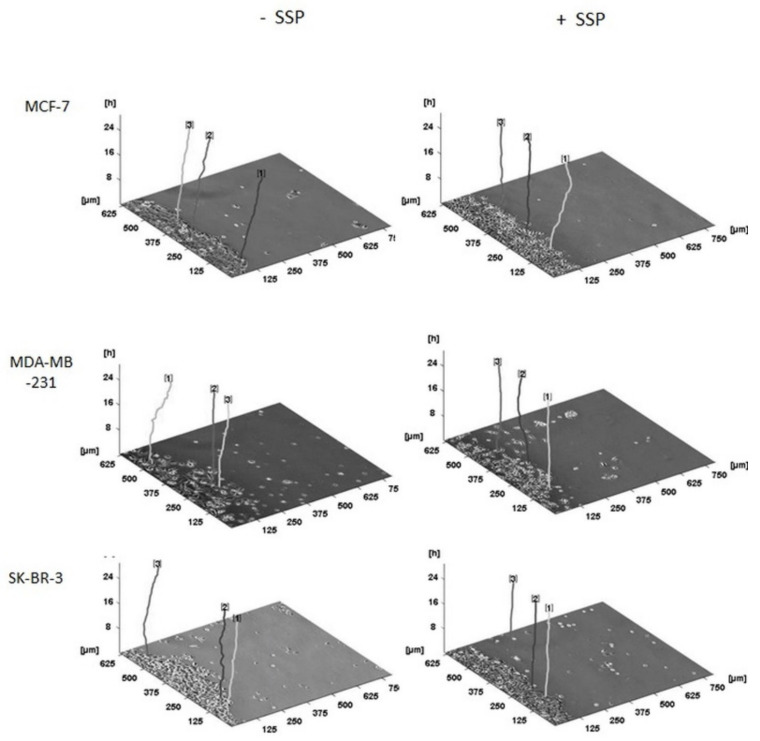
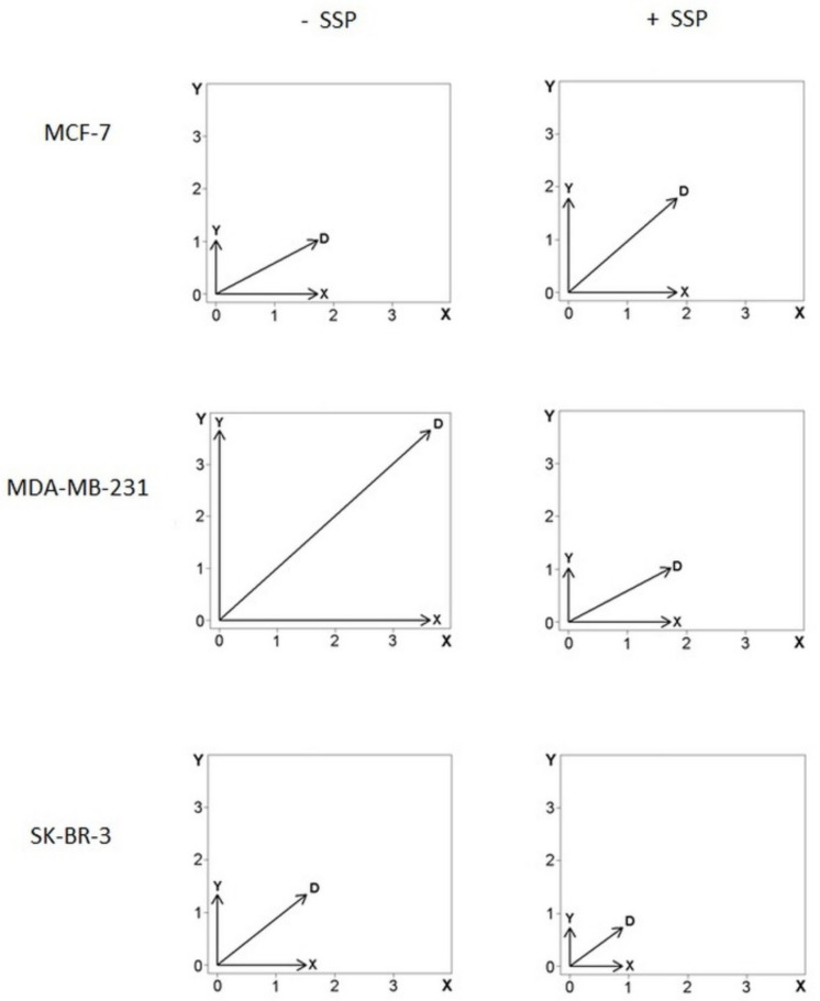
Results: Application of staurosporine induced the migration of single MCF-7 cells but inhibited collective cell migration. With the exception of low-density SK-BR-3 cells, staurosporine induced the generation of immobile flattened giant cells. Video time-lapse analysis revealed that within the borderline of cell collectives, staurosporine reduced the velocity of individual MDA-MB-231 and SK-BR-3, but not of MCF-7 cells. In individual MCF-7 cells, mainly the directionality of migration became disturbed, which led to an increased migration rate parallel to the borderline, and hereby to an inhibition of the migration of the cell collective as a total. Moreover, the application of staurosporine led to a transient activation of ERK1/2 in all cell lines.

Conclusion: Dependent on the context (single versus collective cells), a drug may induce opposite effects in the same cell line.

Keywords: breast carcinoma; cell migration; invasion; staurosporine; yin-yang effect.


Conflict of interest statement

The authors declare no conflict of interest.

Figures

Figure 1
Figure 1
Single-cell migration of breast carcinoma cells on plastic (PL), fibronectin (FN), or laminin (LN) surfaces as revealed by video time-lapse analysis. Histogram shows the velocity of breast carcinoma cells that were analysed for 24 h (given in µm per h + SD). Tracing of the migratory paths was accomplished with the software “Image J” and “CellTracker”. Per cell line and substratum, at least 20 cells were analysed. Selected micrographs show breast carcinoma cells that had been cultivated for 24 h on a plastic (PL), a fibronectin (FN), or a laminin (LN) substratum. Micrographs of identical sections at the beginning (T0) and after 24 h (T24) of the culture period are shown (bar, 50 μm). Notice that in some combinations, such as MCF-7 or SK-BR-3 cells cultivated on PL, almost identical positions of immobile but proliferating cells are present, whereas considerable but variable cell movements occur in other combinations, such as MCF-7 or MDA-MB-231 cells cultivated on LN (also reflected by the large SD values in the histogram).
Figure 2
Figure 2
Collective cell migration of breast carcinoma cells cultivated on plastic surfaces. (A) Histogram shows the increase in the diameter (given in μm per h + SD) of circular areas covered with a confluent layer of breast carcinoma cells after three days in culture (for MCF-7 cells, see subfigure (B)). At least twelve circular areas were measured per experiment and at least three independent experiments per cell line were performed. (B) Low-magnification micrographs of representative circular areas covered with a confluent layer of breast carcinoma cells at the onset of the experiment (T0) and three days later (T72) (bar, 200 μm). (C) High-magnification micrographs of the borderline of circular areas covered with confluent layers of breast carcinoma cells at the beginning of the experiment and one day later (T24) (bar, 60 μm). For MCF-7 cells, big arrows mark the changing position of a single cell that becomes integrated in the cell collective. Small arrows mark the constant position of a small cell cluster outside the cell collective.
Figure 3
Figure 3
Single-cell migration of breast carcinoma cells on plastic (PL), fibronectin (FN), or laminin (LN) surfaces in the absence or presence of 50 nM of SSP, as revealed by video time-lapse analysis. (A) Histogram shows the velocity of breast carcinoma cells that were analysed for 24 h (given in μm per h + SD). With the exception of MCF-7 cells on LN and MDA-MB-231 cells on PL, the differences between untreated and SSP-treated cells are statistically significant, as determined by Student’s t-test. ☠: MDA-MB-231 cells on FN did not survive SSP treatment. (B) Selected micrographs of breast carcinoma cells that had been cultivated for 24 h in the presence of 50 nM of SSP on a PL, FN, or LN substratum. Micrographs of identical sections at the onset of the experiment (T0) and 24 h later (T24) are shown. Notice the changed positions of cells of MCF-7 cells cultivated on PL or FN. In each case, the position of two cells is indicated by small or large arrows (bar, 40 μm). Notice the presence of immobile flattened MDA-MB-231 and SK-BR-3 cells cultivated on LN.
Figure 4
Figure 4
Western blot analysis of ERK1/2 activation in breast carcinoma cells. (A) Breast carcinoma cells were cultured in DMEM, 10% FCS, and directly solubilised (0 h) or solubilised after incubation with 50 nM of SSP for the indicated time spans. (B) Breast carcinoma cells were directly solubilised (control) or, before solubilisation, treated for the indicated time spans with the MEK inhibitor U0126 (20 μM) or for 3 h with 50 nM of SSP either in the absence or presence of 20 μM of U0126. α-Tubulin was used as a loading control. Numbers show fold change compared to controls (set as “1.0”).
Figure 5
Figure 5
Collective cell migration of breast carcinoma cells cultivated on plastic surfaces in the absence or presence of 50 nM of SSP. (A) Histogram shows the relative SSP-provoked inhibition of the migration rate (given in %), i.e., the change in the diameter of a circular area covered with a confluent layer of breast carcinoma cells after three days of culture (for MCF cells, see subfigure (B)). Per experiment, at least twelve circular areas were measured and at least three independent experiments per cell line were performed. The dashed line represents the migration rate of untreated cells that was artificially set as 100%. (B) Micrographs show the borderline of a circular area covered with a confluent layer of breast carcinoma cells after a cultivation period of 24 h in the absence or presence of 50 nM of SSP (bar, 60 μm). Dashed lines mark the position of the border at the beginning of the experiment.
Figure 6
Figure 6
Two-dimensional analysis of the migration pattern of collective border breast carcinoma cells. Collective breast carcinoma cells were allowed to migrate for 24 h in the absence (-SSP) or presence (+SSP) of 50 nM of SSP. The paths of at least 40 carcinoma cells derived from two independent experiments were recorded and integrated into a 2D coordinate as a series of coordinates. With the help of especially designed R-scripts, the different starting points of all cells at T0 were superimposed in the intercept of the “zero” lines in all subfigures, and then the corresponding paths (shown in light grey) were integrated into the 2D coordinate system. Thereby, the paths were reoriented such that the main direction of migration on the abscissa was oriented to the right (see Figure 5B as a comparison). Each black curved line represents a “summarised path” which was calculated for each time point for the position of all individual cells analysed at a certain time point (total time span 24 h, divided from T0 to T72 in 20 min intervals). The individual coordinates of the “summarised path” are based on box and whisker plots for each time point. Hereby, on the X-coordinate, the medians of all 20 min intervals for all cells are presented, whereas on the Y-coordinate, the corresponding lower and upper whisker values or the lower Q25 and upper Q75 quartile values are provided. This set of individual coordinates represented by the summarised paths allows the generation of regression lines. The raise of such regression lines can vary between 0 and 90 degrees, or 0 and –90 degrees, respectively. The angles which can thereby be generated express borders defined by either the lower and upper whiskers (wider angles) and encompass the majority of all path segments, or the lower Q25 and upper Q75 quartile (narrower angles) and encompass 50% of all path segments. Numbers at the X- and Y-axes represent μm.
Figure 7
Figure 7
Three-dimensional migration pattern of selected collective borderline breast carcinoma cells. Collective breast carcinoma cells were allowed to migrate for 24 h in the absence (–SSP) or presence (+SSP) of 50 nM of SSP. The main direction of migration is oriented to the right. The time-dependent (z-axis) paths of three representative carcinoma cells (located in the borderline of the cell collective) per cell line and treatment are shown. For all paths, the endpoint on the z-axis is located at the 24 h position. Thus, the total length of the individual lines may differ, based on a variation of the curvature of the paths. Processing of the primary data was performed with the program “CellTracker” that allows the documentation only in pixel format. One pixel was then converted to 1.25 μm. Numbers at the top of the paths were automatically set by the program.
Figure 8
Figure 8
Vector diagrams of cell velocities from individual collective border breast carcinoma cells. Data were summarised from cells that were analysed as shown in Figure 6. The X-axis is oriented to the main direction and represents the changes of the mean velocity given by the x-coordinates (μm per h) oriented into the main direction of the overall migration of the collective (see text for details and Figure 5 for orientation). The Y-axis represents the changes of the mean velocity given by the y-coordinates (μm per h). The diagonal, D, represents the sum of the vectors X and Y with the vector’s magnitude defined by ||D|| = (sqrt(X2 + Y2)) in μm per h. For details, see text.

References

    1. Bill R., Christofori G. The relevance of EMT in breast cancer metastasis: Correlation or causality? FEBS Lett. 2015;589:1577–1587. doi: 10.1016/j.febslet.2015.05.002. - DOI - PubMed
    1. Yilmaz M., Christofori G. Mechanisms of motility in metastasizing cells. Mol. Cancer Res. 2010;8:629–642. doi: 10.1158/1541-7786.MCR-10-0139. - DOI - PubMed
    1. Lobastova L., Kraus D., Glassmann A., Khan D., Steinhauser C., Wolff C., Veit N., Winter J., Probstmeier R. Collective cell migration of thyroid carcinoma cells: A beneficial ability to override unfavourable substrates. Cell Oncol. 2017;40:63–76. doi: 10.1007/s13402-016-0305-5. - DOI - PubMed
    1. Friedl P., Wolf K. Plasticity of cell migration: A multiscale tuning model. J. Cell Biol. 2010;188:11–19. doi: 10.1083/jcb.200909003. - DOI - PMC - PubMed
    1. Odenthal J., Takes R., Friedl P. Plasticity of tumor cell invasion: Governance by growth factors and cytokines. Carcinogenesis. 2016;37:1117–1128. doi: 10.1093/carcin/bgw098. - DOI - PubMed

Publication types