. 2021 Nov 4;22(21):11967.
doi: 10.3390/ijms222111967.

Senolytic Therapy for Cerebral Ischemia-Reperfusion Injury

Affiliations

Senolytic Therapy for Cerebral Ischemia-Reperfusion Injury

Songhyun Lim et al. Int J Mol Sci. .

Abstract

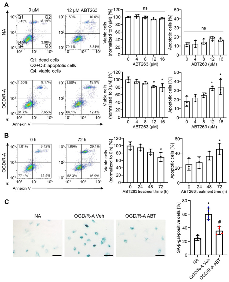
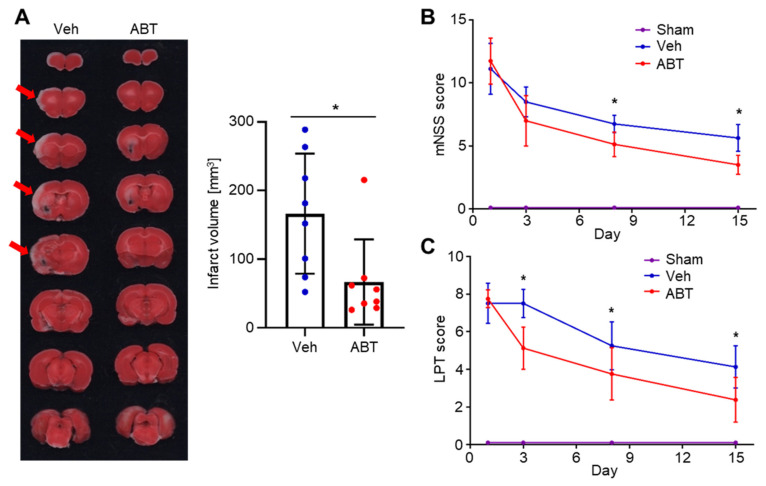
Ischemic stroke is one of the leading causes of death, and even timely treatment can result in severe disabilities. Reperfusion of the ischemic stroke region and restoration of the blood supply often lead to a series of cellular and biochemical consequences, including generation of reactive oxygen species (ROS), expression of inflammatory cytokines, inflammation, and cerebral cell damage, which is collectively called cerebral ischemia-reperfusion (IR) injury. Since ROS and inflammatory cytokines are involved in cerebral IR injury, injury could involve cellular senescence. Thus, we investigated whether senolytic therapy that eliminates senescent cells could be an effective treatment for cerebral IR injury. To determine whether IR induces neural cell senescence in vitro, astrocytes were subjected to oxygen-glucose deprivation/reoxygenation (OGD/R). OGD/R induced astrocyte senescence and senescent cells in OGD/R-injured astrocytes were effectively eliminated in vitro by ABT263, a senolytic agent. IR in rats with intraluminal middle cerebral artery occlusion induced cellular senescence in the ischemic region. The senescent cells in IR-injured rats were effectively eliminated by intravenous injections of ABT263. Importantly, ABT263 treatment significantly reduced the infarct volume and improved neurological function in behavioral tests. This study demonstrated, for the first time, that senolytic therapy has therapeutic potential for cerebral IR injury.

Keywords: ABT263; astrocyte; inflammation; ischemia-reperfusion injury; ischemic stroke; senescence; senolytic therapy.


Conflict of interest statement

The authors declare no conflict of interest.

Figures

Figure 1
Figure 1
Rat astrocyte senescence induced in vitro by oxygen-glucose deprivation/reoxygenation (OGD/R). (A) Viability and death of normal astrocytes (NA) and OGD/R-injured astrocytes (OGD/R-A), as evaluated by 3-(4,5-dimethylthiazol-2-yl)-2,5-diphenyltetrazolium bromide (MTT) assay and lactate dehydrogenase (LDH) release assay, respectively. OGD/R injury showed no cytotoxicity to rat cortex astrocytes. ns = not significant. n = 4 per group. (B) Representative images and quantification of senescence-associated beta-galactosidase (SA-β-gal) staining (green) after 4 h of OGD/R injury. OGD/R injury induced senescence in rat cortex astrocytes. Scale bars, 100 µm. n = 4 per group. (A,B) Data are presented as mean ± SD. * p < 0.05. Mann–Whitney test was used for statistical analysis.
Figure 2
Figure 2
Selective removal of senescent astrocytes by ABT263 in vitro. (A) Flow cytometric plots of normal astrocytes (NA) and OGD/R-injured astrocytes (OGD/R-A), showing selective induction of OGD/R-A apoptosis by ABT263. (B) Flow cytometric plots of OGD/R-A treated with 12 µM ABT263 for different periods of time. To effectively remove senescent astrocytes, the duration period of ABT263 treatment should be at least 72 h. (C) SA-β-gal staining of NA and OGD/R-A treated with either vehicle or 12 µM ABT263 for 96 h, showing removal of senescent cells of OGD/R-A. Veh indicates vehicle treatment, and ABT indicates ABT263 treatment. Scale bars, 100 µm. n = 4 per group. * p < 0.05 compared to NA, # p < 0.05 compared to OGD/R-A Veh. (A,B) n = 3 per group. * p < 0.05 compared to 0 µM or 0 h. (AC) Data are presented as mean ± SD. One-way ANOVA with Tukey’s post-test was used for statistical analysis.
Figure 3
Figure 3
Removal of senescent cells and attenuation of inflammation in ischemic stroke lesion by ABT263 in middle cerebral artery occlusion (MCAO) ischemia-reperfusion rat model. MCAO rats were treated with intravenous injection of either vehicle (Veh) or ABT263 (ABT). The sham group did not receive MCAO. Immunostaining for (A) p16INK4a, (B) NOS2, (C) MPO and (D) GFAP in the ischemic stroke lesion of MCAO rats on day 4. (AD) Scale bars, 100 µm. n = 6 per group. Data are presented as mean ± SD. * p < 0.05 compared to Sham, # p < 0.05 compared to Veh. One-way ANOVA with Tukey’s post-test was used for statistical analysis.
Figure 4
Figure 4
Therapeutic effect of ABT263 in MCAO ischemia-reperfusion rat model. MCAO rats were treated with intravenous injections of either vehicle (Veh) or ABT263 (ABT). The sham group did not receive MCAO. (A) 2,3,5-triphenyltetrazolium chloride (TTC) staining of serial sections of the brain and quantification of the cerebral infarct volume on day 4. The ABT263 treatment group had significantly smaller infarct volume compared to the Veh group. Arrows indicate cerebral infarct. n = 8 per group. * p < 0.05. Data are presented as mean ± SD. Mann–Whitney test was used for statistical analysis. Brain functional recoveries of MCAO rats evaluated by (B) the limb placing test (LPT) and (C) modified neurological severity score (mNSS). The mNSS tests and the limb-placing tests showed that ABT263 treatment significantly improved neurological functions after MCAO. n = 6 per group. Data are presented as mean ± SD. * p < 0.05 compared to Veh or Sham. Two-way repeated measures ANOVA was used for statistical analysis.

References

    1. Grysiewicz R.A., Thomas K., Pandey D.K. Epidemiology of ischemic and hemorrhagic stroke: Incidence, prevalence, mortality, and risk factors. Neurol. Clin. 2008;26:871–895. doi: 10.1016/j.ncl.2008.07.003. - DOI - PubMed
    1. Gorelick P.B. The global burden of stroke: Persistent and disabling. Lancet Neurol. 2019;18:417–418. doi: 10.1016/S1474-4422(19)30030-4. - DOI - PubMed
    1. Venketasubramanian N., Yoon B.W., Pandian J., Navarro J.C. Stroke epidemiology in south, east, and south-east asia: A review. J. Stroke. 2017;19:286. doi: 10.5853/jos.2017.00234. - DOI - PMC - PubMed
    1. Lo E.H., Dalkara T., Moskowitz M.A. Mechanisms, challenges and opportunities in stroke. Nat. Rev. Neurosci. 2003;4:399–414. doi: 10.1038/nrn1106. - DOI - PubMed
    1. Nour M., Scalzo F., Liebeskind D.S. Ischemia-reperfusion injury in stroke. Interv. Neurol. 2012;1:185–199. doi: 10.1159/000353125. - DOI - PMC - PubMed

LinkOut - more resources