. 2021 Nov 5;22(21):11974.
doi: 10.3390/ijms222111974.

A Potential New Role for Zinc in Age-Related Macular Degeneration through Regulation of Endothelial Fenestration

Affiliations

A Potential New Role for Zinc in Age-Related Macular Degeneration through Regulation of Endothelial Fenestration

Fiona Cunningham et al. Int J Mol Sci. .

Abstract

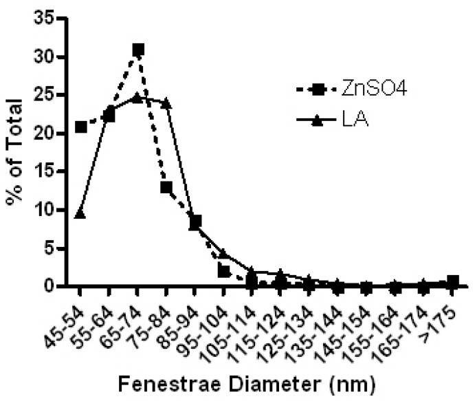
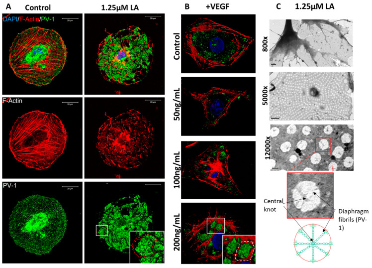
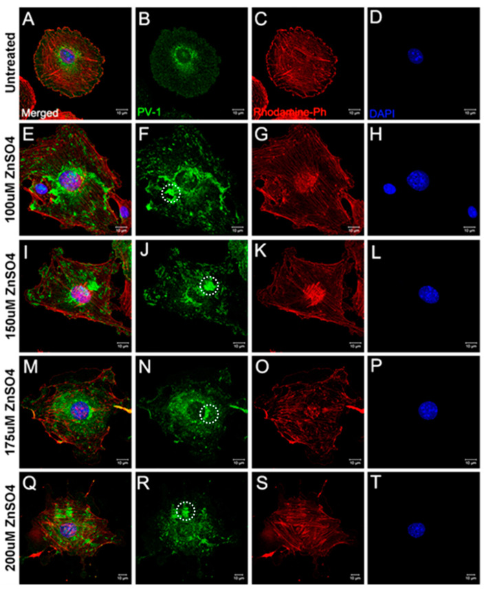
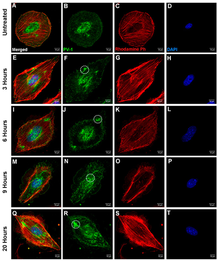
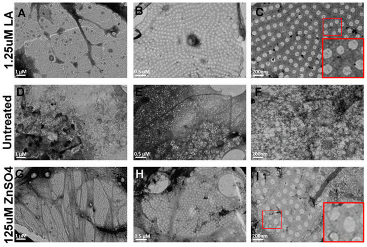
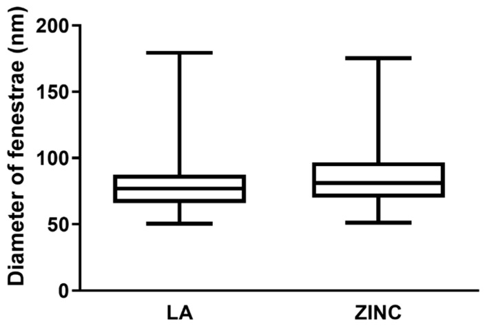
Age-related macular degeneration (AMD) is a common blinding disease in the western world that is linked to the loss of fenestration in the choriocapillaris that sustains the retinal pigment epithelium and photoreceptors in the back of the eye. Changes in ocular and systemic zinc concentrations have been associated with AMD; therefore, we hypothesized that these changes might be directly involved in fenestrae formation. To test this hypothesis, an endothelial cell (bEND.5) model for fenestrae formation was treated with different concentrations of zinc sulfate (ZnSO4) solution for up to 20 h. Fenestrae were visualized by staining for Plasmalemmal Vesicle Associated Protein-1 (PV-1), the protein that forms the diaphragms of the fenestrated endothelium. Size and distribution were monitored by transmission electron microscopy (TEM). We found that zinc induced the redistribution of PV-1 into areas called sieve plates containing ~70-nm uniform size and typical morphology fenestrae. As AMD is associated with reduced zinc concentrations in the serum and in ocular tissues, and dietary zinc supplementation is recommended to slow disease progression, we propose here that the elevation of zinc concentration may restore choriocapillaris fenestration resulting in improved nutrient flow and clearance of waste material in the retina.

Keywords: PV-1; age-related macular degeneration; choroid; fenestration; zinc.


Conflict of interest statement

While SC is working for Nestlé, this work was done as part of her Ph.D. thesis, and therefore, no conflict of interest exists.

Figures

Figure A1
Figure A1
Distribution of fenestrae diameter induced by zinc or LA. Graphical representation of data in Table A1. The distribution of fenestrae diameters was similar in LA- and ZnSO4-treated cells.
Figure 1
Figure 1
bEND.5 cells have the potential to become fenestrated. (A). BEND.5 cells treated with 1.25 μM of LA had PV-1 (green) and actin (red) redistribution to sieve plates as compared to control non-treated cells (sieve plate indicated by white box and red dotted line). (B). VEGF-induced sieve plate formation could also be stimulated in bEND.5 cells as illustrated by the rearrangement of PV-1 (green) and the actin cytoskeleton (red) (white box and dotted line indicate sieve plate formation in 200 ng/mL of VEGF-treated bEND.5 cells). (C). TEM showed uniform distribution of fenestrae (800× magnification, scale bar: 1 µM; 5000× magnification, scale bar: 0.5 µM). At high magnification, fenestrae diaphragm fibrils and central knots could be visualized (12,000× magnification, scale bar: 200 nm) as highlighted in the accompanying diagram.
Figure 2
Figure 2
Sieve plate formation induced by zinc sulfate. 100–200 μM of ZnSO4 induced PV-1 redistribution (green) and sieve plate formation (F,J,N,R), examples indicated by white dotted circles) compared to untreated control cells (B). However, there were no significant actin (red) cytoskeletal changes (C,G,K,O,S) or degradation of the nucleus (blue) (D,H,L,P,T). (A,E,I,M,Q) show the images after merging the different fluorescence channels.
Figure 3
Figure 3
Time-dependent sieve plate formation induced by zinc. When compared to control (B) 125 μM ZnSO4 induced PV-1 (green) rearrangement as early as 3 h after treatment initiation (F); however, sieve plate formation became clearly apparent in cells treated for 9 or more hours (N,R). Sieve plate formation is indicated by white dotted circles (F,J,N,R). There were no significant actin (red) cytoskeletal changes (C,G,K,O,S) or degradation of the nucleus (blue) (D,H,L,P,T). (A,E,I,M,Q) show the images after merging the different fluorescence channels.
Figure 4
Figure 4
Zinc-induced fenestrae formation was examined by TEM. Fenestrae formation induced by LA (AC) was used as a positive control for these observations. In untreated cells, there was some spontaneous fenestrae formation; however, these lacked uniformity and diaphragm organization (DF). As compared to LA-treated cells, zinc induced comparatively smaller sieve plates without major cytoskeletal rearrangements (G,H). However, zinc-induced fenestrae were uniform and exhibited typical morphology, including diaphragm formation (I), like those induced by LA (C). (Scale bar, (A,D,G) = 1 micron; (B,E,H) = 0.5 micron; (C,F,H) = 200 nm).
Figure 5
Figure 5
Size distribution of fenestrae diameter induced by zinc or LA. The distribution of fenestrae diameters was similar in LA and ZnSO4 treated cells. A total of 775 fenestrae from LA-treated cells and 393 fenestrae from ZnSO4-treated cells were assessed from 3 independent experiments.

References

    1. de Jong P.T.V.M. Age-Related Macular Degeneration. N. Engl. J. Med. 2006;355:1474–1485. doi: 10.1056/NEJMra062326. - DOI - PubMed
    1. Al-Zamil W., Yassin S. Recent developments in age-related macular degeneration: A review. Clin. Interv. Aging. 2017;12:1313–1330. doi: 10.2147/CIA.S143508. - DOI - PMC - PubMed
    1. Vishwanathan R., Chung M., Johnson E.J. A Systematic Review on Zinc for the Prevention and Treatment of Age-Related Macular Degeneration. Investig. Ophthalmol. Vis. Sci. 2013;54:3985–3998. doi: 10.1167/iovs.12-11552. - DOI - PubMed
    1. Age-Related Eye Disease Study Research Group A Randomized, Placebo-Controlled, Clinical Trial of High-Dose Supplementation with Vitamins C and E, Beta Carotene, and Zinc for Age-Related Macular Degeneration and Vision Loss: AREDS Report No. 8. Arch. Ophthalmol. 2001;119:1417–1436. doi: 10.1001/archopht.119.10.1417. - DOI - PMC - PubMed
    1. van Leeuwen R., Boekhoorn S., Vingerling J.R., Witteman J.C.M., Klaver C.C.W., Hofman A., de Jong P.T.V.M. Dietary Intake of Antioxidants and Risk of Age-Related Macular Degeneration. JAMA. 2005;294:3101–3107. doi: 10.1001/jama.294.24.3101. - DOI - PubMed

MeSH terms