Review
. 2021 Nov 5;22(21):11976.
doi: 10.3390/ijms222111976.

Single-Cell RNA Sequencing Reveals Heterogeneity and Functional Diversity of Lymphatic Endothelial Cells

Affiliations
Review

Single-Cell RNA Sequencing Reveals Heterogeneity and Functional Diversity of Lymphatic Endothelial Cells

Hannah den Braanker et al. Int J Mol Sci. .

Abstract

Lymphatic endothelial cells (LECs) line the lymphatic vasculature and play a central role in the immune response. LECs have abilities to regulate immune transport, to promote immune cell survival, and to cross present antigens to dendritic cells. Single-cell RNA sequencing (scRNA) technology has accelerated new discoveries in the field of lymphatic vascular biology. This review will summarize these new findings in regard to embryonic development, LEC heterogeneity with associated functional diversity, and interactions with other cells. Depending on the organ, location in the lymphatic vascular tree, and micro-environmental conditions, LECs feature unique properties and tasks. Furthermore, adjacent stromal cells need the support of LECs for fulfilling their tasks in the immune response, such as immune cell transport and antigen presentation. Although aberrant lymphatic vasculature has been observed in a number of chronic inflammatory diseases, the knowledge on LEC heterogeneity and functional diversity in these diseases is limited. Combining scRNA sequencing data with imaging and more in-depth functional experiments will advance our knowledge of LECs in health and disease. Building the case, the LEC could be put forward as a new therapeutic target in chronic inflammatory diseases, counterweighting the current immune-cell focused therapies.

Keywords: inflammation; lymph nodes; lymphatic endothelial cells; lymphatics; single cell RNA-sequencing.


Conflict of interest statement

The authors declare no conflict of interest.

Figures
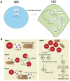
Figure 1
Figure 1
Lymphatic endothelial cell and lymph node embryonic development. (A) In embryonic development, lymphatic endothelial cells (LECs) mostly transdifferentiate from venous endothelial cells (VECs). Prospero homeobox protein 1 (Prox-1) is a master regulator of lymphatic fate. Homeobox d 8 (Hoxd8) can activate Prox-1 expression, and Prox-1 cooperates with Nuclear Receptor Subfamily2 Group F Member 2 (Nr2f2) to drive expression of other key peripheral LEC molecules, such as podoplanin(Pdpn), lymphatic vessel endothelial hyaluron receptor 1 (Lyve-1), chemokine (C-C motif) ligand 21 (Ccl21), and vascular endothelial growth factor receptor 3 (Vegfr3). Vegfr3 is also regulated by transcription factor T-box 1 (Tbx1) and can activate other transcription factors, such as Maf. Single cell RNA sequencing (scRNA-seq) data also suggested transcription factors, such as ETS Transcription Factor 3 (Elk3), or other protein coding RNAs, such as Profilin 1 (Pfn1) and AE binding protein 1 (Aebp1), to be involved in lymphatic development, but their function is not yet clear. (B) Lymph node (LN) development follows several steps: (1) mesenchymal cells secrete chemokine (C-X-C motif) ligand 13 (CXCL13) to attract lymphoid tissue inducer (LTi) cells. (2) LTi cells cluster together and activate receptor activator nuclear factor κ B (RANK) and RANK ligand (RANKL) signaling. (3) RANK-RANKL signaling increases lymphotoxin α1β2 (LT1β2), which activates lymphotoxin beta receptor (LTβR) on mesenchymal cells responding with more cytokine signaling and attracting more LTi cells. (4) Mesenchymal cells differentiate into stromal cell subsets of the LN, and LTi differentiate or attract LECs. Trajectory inference of scRNA-seq data showed that there are two developmental paths of the LN LECs.
Figure 2
Figure 2
Overview of the different lymph node lymphatic endothelial cells and peripheral lymphatic endothelial cell subpopulations. The lymph node (LN) consists of multiple sinuses to transport lymph and immune cells from the afferent lymphatic vessels to the efferent lymphatic vessel. Immune cells arrive in the subcapular sinus (SCS) and migrate over the floor of the SCS, lined by floor LECs, to the LN cortex. The SCS is a narrow sinus, and it is close to the LN capsule. The LN capsule side of the SCS is lined by ceiling LECs. In the LN cortex, immune cells also arrive via high endothelial venules (HEVs). Egress of immune cells from the cortex is via the cortical sinuses (CS) lined by cortical LECs. These cortical sinuses drain in the medulla. Medullary sinuses (MS), lined by medullary LECs, transport lymph and immune cells to the efferent lymphatic vessel. LECs lining the different sinuses were found to be transcriptionally different and to have a range of different functions.
Figure 3
Figure 3
Inflammatory changes in lymph node lymphatic endothelial cells and peripheral lymphatic endothelial cells. (A) Predominantly, lymphatic endothelial cells (LECs) lining the floor of the subcapular sinus (SCS) (floor LECs) come into contact with immune cells of inflamed tissue. Floor LECs can rapidly upregulate the expression of chemokines, such as chemokine (C-C motif) ligand 20 (CCL20). Additionally, molecules such as Annexin A2 (ANXA-2) and cluster of differentiation 200 (CD200) are upregulated. The function of these molecules in the inflamed LN needs to be investigated. (B) Oxidized low-density lipoprotein (LDL) can downregulate transcripts like Prospero Homeobox Protein 1 (PROX-1), Podoplanin (PDPN), and Vascular Endothelial Growth Factor Receptor 3 (VEGFR3), which are key LEC markers. CD36, a fatty acid transporter, and CCL21 are upregulated. Single cell RNA sequencing (ScRNA-seq) data on inflammatory changes by cytokines or immune cells have not yet been investigated.

References

    1. Wiig H., Swartz M.A. Interstitial Fluid and Lymph Formation and Transport: Physiological Regulation and Roles in Inflammation and Cancer. Physiol. Rev. 2012;92:1005–1060. doi: 10.1152/physrev.00037.2011. - DOI - PubMed
    1. Petrova T.V., Koh G.Y. Biological functions of lymphatic vessels. Science. 2020;369:eaax4063. doi: 10.1126/science.aax4063. - DOI - PubMed
    1. Oliver G., Kipnis J., Randolph G.J., Harvey N.L. The Lymphatic Vasculature in the 21st Century: Novel Functional Roles in Homeostasis and Disease. Cell. 2020;182:270–296. doi: 10.1016/j.cell.2020.06.039. - DOI - PMC - PubMed
    1. Tewalt E.F., Cohen J.N., Rouhani S.J., Engelhard V.H. Lymphatic endothelial cells-key players in regulation of tolerance and immunity. Front. Immunol. 2012;3:305. doi: 10.3389/fimmu.2012.00305. - DOI - PMC - PubMed
    1. Förster R., Braun A., Worbs T. Lymph node homing of T cells and dendritic cells via afferent lymphatics. Trends Immunol. 2012;33:271–280. doi: 10.1016/j.it.2012.02.007. - DOI - PubMed

LinkOut - more resources