. 2021 Nov 5;22(21):11986.
doi: 10.3390/ijms222111986.

Establishment of Human-Induced Pluripotent Stem Cell-Derived Neurons-A Promising In Vitro Model for a Molecular Study of Rabies Virus and Host Interaction

Affiliations

Establishment of Human-Induced Pluripotent Stem Cell-Derived Neurons-A Promising In Vitro Model for a Molecular Study of Rabies Virus and Host Interaction

Thanathom Chailangkarn et al. Int J Mol Sci. .

Abstract

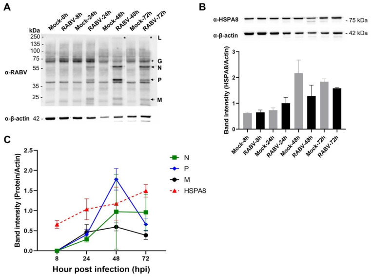
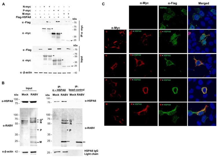
Rabies is a deadly viral disease caused by the rabies virus (RABV), transmitted through a bite of an infected host, resulting in irreversible neurological symptoms and a 100% fatality rate in humans. Despite many aspects describing rabies neuropathogenesis, numerous hypotheses remain unanswered and concealed. Observations obtained from infected primary neurons or mouse brain samples are more relevant to human clinical rabies than permissive cell lines; however, limitations regarding the ethical issue and sample accessibility become a hurdle for discovering new insights into virus-host interplays. To better understand RABV pathogenesis in humans, we generated human-induced pluripotent stem cell (hiPSC)-derived neurons to offer the opportunity for an inimitable study of RABV infection at a molecular level in a pathologically relevant cell type. This study describes the characteristics and detailed proteomic changes of hiPSC-derived neurons in response to RABV infection using LC-MS/MS quantitative analysis. Gene ontology (GO) enrichment of differentially expressed proteins (DEPs) reveals temporal changes of proteins related to metabolic process, immune response, neurotransmitter transport/synaptic vesicle cycle, cytoskeleton organization, and cell stress response, demonstrating fundamental underlying mechanisms of neuropathogenesis in a time-course dependence. Lastly, we highlighted plausible functions of heat shock cognate protein 70 (HSC70 or HSPA8) that might play a pivotal role in regulating RABV replication and pathogenesis. Our findings acquired from this hiPSC-derived neuron platform help to define novel cellular mechanisms during RABV infection, which could be applicable to further studies to widen views of RABV-host interaction.

Keywords: human-induced pluripotent stem cells; in vitro model; neurons; proteomics analysis; rabies virus; virus–host interaction.


Conflict of interest statement

The authors declare no conflict of interest. The funders had no role in the design of the study; in the collection, analyses, or interpretation of data; in the writing of the manuscript; or in the decision to publish the results.

Figures

Figure 1
Figure 1
Generation and characterization of hiPSC-derived NPCs, neurons, and astrocytes and their expression of specific markers by IFA. (A) Schematic diagram summarizing the derivation of NPCs, neurons, and astrocytes from human iPSCs. (B) Undifferentiated NPCs were stained with mouse α-Nestin, rabbit α-Pax6, rabbit α-Musashi1, and rabbit α-Sox1 as primary antibodies. Goat α-mouse IgG Texas Red and goat α-rabbit IgG Alexa Fluor® 488 were used as secondary antibodies. Scale bars = 20 µm. (C) Neurons were stained with mouse α-β-tubulin III and rabbit α-MAP2. Astrocytes were stained with mouse α-GFAP and rabbit α-S100β as primary antibodies. Goat α-mouse IgG Texas Red, α-rabbit IgG TRITC and α-rabbit IgG Alexa Fluor® 488 were used as the secondary antibodies. Scale bars = 50 µm.
Figure 2
Figure 2
Transcriptomic and gene expression profiles of hiPSC-derived NPCs, neurons, and astrocytes. (A) Principal component analysis plot displaying all 12 RNA libraries along PC1 and PC2, which described 89% and 7% of the variability within the expression data set. (B) Hierarchical clustering analysis of all 12 RNA libraries based on Euclidian distances using DESeq2 package. (C) Gene expression profile of hiPSC-derived NPCs, neurons, and astrocytes quantified by real-time RT-PCR.
Figure 3
Figure 3
hiPSC-derived NPCs, neurons, and astrocytes are permissive to rabies infection. (A) All cell types were infected with RABV-TH at MOI of 0.5 for 72 h. The cultures were co-stained with horse α-rabies serum and mouse α-Nestin, MAP2, or S100β antibodies for NPCs, neurons, and astrocytes, respectively. The cells were observed by fluorescence microscopy. Scale bars = 50 µm. (B) Cells were infected with RABV-TH at MOI of 0.5. Cell supernatants were collected daily for virus titration in BHK21 cells. Error bars represent the standard deviation of virus titers (TCID50/mL) from three independent experiments.
Figure 4
Figure 4
RABV infection induced apoptosis in hiPSC-derived NPC but not neurons and astrocytes. Cells were infected with RABV-TH at MOI 0.5. NPCs were maintained in NPC media (undifferentiated stage) or in NPC media with the withdrawal of FGF2 (NPCs undergoing neuronal differentiation). Neurons and astrocytes were maintained in NPC media without FGF2, and astrocyte media supplemented with FBS. To examine apoptosis in each cell type by flow cytometry, (A) NPCs at different stages included undifferentiated NPCs, and NPCs undergoing neuronal differentiation were harvested at 72 hpi and 5 days post infection (dpi), respectively, and (B) neurons and astrocytes were harvested at 72 hpi. Percentages of apoptotic cells shown are representative of triplicate. Error bars represent means ± SEM. Data were analyzed by an unpaired-t test. ns, p > 0.05; * p < 0.05; ** p < 0.01.
Figure 5
Figure 5
Schematic diagram of proteomic analysis and differential expression protein (DEP) identification in RABV-infected hiPSC-derived neurons. (A) Schematic workflows of sample preparation and bioinformatic analysis. (B) K-means clustering demonstrated 16 clusters of expressed proteins changing in RABV-infected neurons at 8, 24, 48, and 72 hpi.
Figure 6
Figure 6
Hierarchical clustering of the proportion of DEPs among proteins annotated with each heat shock-related and chaperone function. Euclidean metric and complete linkage were used.
Figure 7
Figure 7
Protein–protein interaction network (PPI) of HSPA8 using STRING database. The edges represented in the full network indicate both functional and physical protein associations. Network statistics—number of nodes: 31, number of edges: 280, average node degree: 18.1, average local clustering coefficient: 0.933, expected number of edges: 73, PPI enrichment p-value: <1.0 × 10⁻¹⁶.
Figure 8
Figure 8
A dynamic change of RABV and HSPA8 proteins in infected hiPSC-derived neurons. Protein samples of mock and RABV-infected neurons were subjected to Western blot analysis. The membranes were probed with (A) horse α-RABV serum and (B) mouse α-human HSPA8 antibodies to detect RABV and HSPA8 proteins, respectively. β-actin was used as the internal loading control. (C) The intensity of RABV N, P, M, and HSPA8 in the infection group was normalized with β-actin using an image analysis program. * Note that RABV G and L proteins were excluded from the analysis due to nonspecific bands presented at the same size as RABV G, masking the RABV G and faint bands of RABV L. Error bars represent means of band intensity ± standard deviation of duplicates.
Figure 9
Figure 9
HSPA8 and RABV protein interactions. HSPA8 and RABV proteins’ physical interaction was determined by co-immunoprecipitations of protein samples harvested from HEK293T cells and hiPSC-derived neurons. (A) HEK293T cells were transfected with the plasmids expressing Flag-HSPA8 and RABV Nmyc, Pmyc, and Mmyc. At 48 h post-transfection, cell lysates were collected and prepared for immunoprecipitation using mouse α-myc beads. The eluted proteins were probed with rabbit α-myc and –flag. The arrows specify the presence of each viral protein according to its expected size (N, P, and M proteins at approximately 60, 40, and 25 kDa, respectively). (B) The neurons were infected with RABV-TH at MOI of 0.5. At 48 hpi, the cells were collected and subjected to immunoprecipitation using beads conjugated with mouse α-human HSPA8 antibody. The same samples were incubated with the beads without α-human HSPA8 antibody conjugation as the negative control. * RABV G appears at the same size as a nonspecific band found in a mock sample. ** RABV M might be either masked by the nonspecific band or strongly binds to the beads, despite lysate preclearing and several washing steps. (C) For the colocalization assay, transfected HEK293T cells were fixed with 4% paraformaldehyde and probed with mouse α-myc and rabbit anti-flag as primary antibodies. Goat α-rabbit Alexa Fluor 488 and goat α-mouse Alexa Fluor 647 were used as secondary antibodies.

References

    1. Lafon M. Rabies Virus Receptors. J. Neurovirol. 2005;11:82–87. doi: 10.1080/13550280590900427. - DOI - PubMed
    1. Gillet J.P., Derer P., Tsiang H. Axonal Transport of Rabies Virus in the Central Nervous System of the Rat. J. Neuropathol. Exp. Neurol. 1986;45:619–634. doi: 10.1097/00005072-198611000-00002. - DOI - PubMed
    1. Hemachudha T., Ugolini G., Wacharapluesadee S., Sungkarat W., Shuangshoti S., Laothamatas J. Human Rabies: Neuropathogenesis, Diagnosis, and Management. Lancet Neurol. 2013;12:498–513. doi: 10.1016/S1474-4422(13)70038-3. - DOI - PubMed
    1. Jackson A.C., Reimer D.L. Pathogenesis of Experimental Rabies in Mice: An Immunohistochemical Study. Acta Neuropathol. 1989;78:159–165. doi: 10.1007/BF00688204. - DOI - PubMed
    1. Mitrabhakdi E., Shuangshoti S., Wannakrairot P., Lewis R.A., Susuki K., Laothamatas J., Hemachudha T. Difference in Neuropathogenetic Mechanisms in Human Furious and Paralytic Rabies. J. Neurol. Sci. 2005;238:3–10. doi: 10.1016/j.jns.2005.05.004. - DOI - PubMed