Review
. 2021 Nov 6;22(21):12018.
doi: 10.3390/ijms222112018.

Dental Pulp Stem Cell-Derived Secretome and Its Regenerative Potential

Affiliations
Review

Dental Pulp Stem Cell-Derived Secretome and Its Regenerative Potential

Julia K Bar et al. Int J Mol Sci. .

Abstract

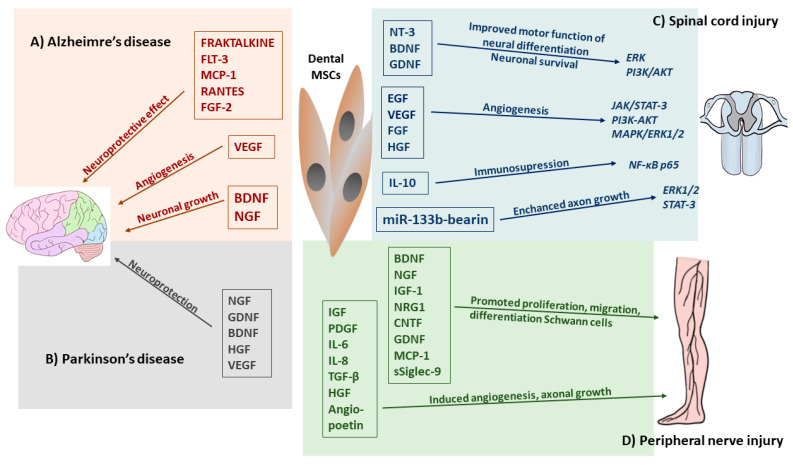
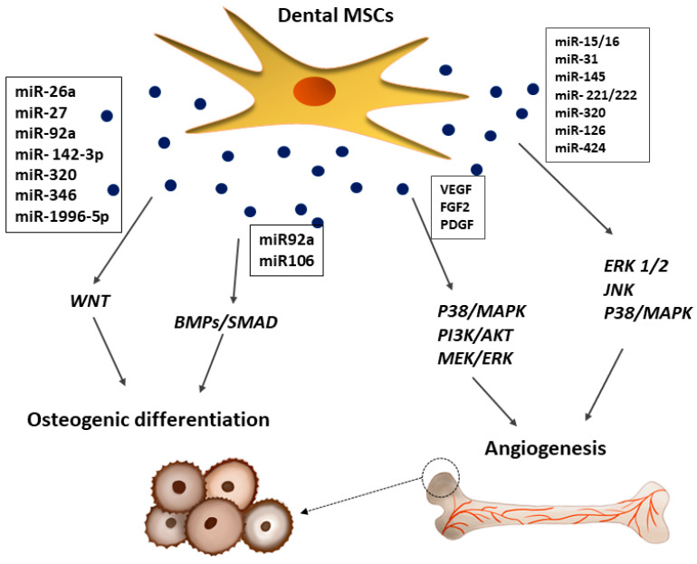
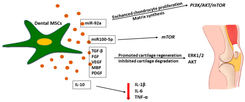
The therapeutic potential of the dental pulp stem (DSC) cell-derived secretome, consisting of various biomolecules, is undergoing intense research. Despite promising in vitro and in vivo studies, most DSC secretome-based therapies have not been implemented in human medicine because the paracrine effect of the bioactive factors secreted by human dental pulp stem cells (hDPSCs) and human exfoliated deciduous teeth (SHEDs) is not completely understood. In this review, we outline the current data on the hDPSC- and SHED-derived secretome as a potential candidate in the regeneration of bone, cartilage, and nerve tissue. Published reports demonstrate that the dental MSC-derived secretome/conditional medium may be effective in treating neurodegenerative diseases, neural injuries, cartilage defects, and repairing bone by regulating neuroprotective, anti-inflammatory, antiapoptotic, and angiogenic processes through secretome paracrine mechanisms. Dental MSC-secretomes, similarly to the bone marrow MSC-secretome activate molecular and cellular mechanisms, which determine the effectiveness of cell-free therapy. Many reports emphasize that dental MSC-derived secretomes have potential application in tissue-regenerating therapy due to their multidirectional paracrine effect observed in the therapy of many different injured tissues.

Keywords: dental stem cells; paracrine effect; regenerative medicine; secretome.


Conflict of interest statement

The authors declare no conflict of interest.

Figures

Figure 1
Figure 1
Composition of bioactive factors release by mesenchymal stem cells.
Figure 2
Figure 2
Dental MSC-derived secretome production and methods of its modification.
Figure 3
Figure 3
Paracrine effect induced by human dental pulp stem cells (hDPSCs) secretome.
Figure 4
Figure 4
Effects of dental MSC-derived secretome in neurodegenerative diseases and central and peripheral nerve injury therapy. (A) Dental MSC-CM such as RANTES, FGF2, and Fractalkine enhanced neuronal cell survival and reduces apoptosis, BDNF, NGF involved in neuronal growth, whereas, VEGF promoted angiogenesis in Alzheimer’s disease. (B) Trophic factors (BDNF, NGF, GDNF, HGC, VEGF) and cytokines (TNF-α, IL-6) released by dental MCS-CM enhanced the regulatory function and reverts the damage sustained by host neurons in Parkinson’s disease. (C) Released by dental MSCs neurotrophic factors promoted neuronal differentiation and survival by activation of Erk, PI3/AKT and growth factors induced angiogenesis through JAK/STAT3, PI3K/AKT MAPK/Erk1/2 signaling pathways. Administration of dental MSC-EVs, miR-133 bearing promoted recovery form SCI by enhancing regeneration of axons through the activation of survival Erk1/2 and STAT-3 signaling pathways in regenerating neurons. Dental MSC-EVs induced immunosuppression at the site of SCI by enhancing production of IL-10 which suppressed neurotoxic A1 astrocytes through the inhibition of NF-κB-p65 signaling pathways. (D) Dental-MCS-derived secretome modulates nerve regeneration of peripheral nerve injury (PNI) by secretion of many neurotrophic factors which promoted survival of neuronal and glial cells, whereas, MCP-1 and sSiglec-9 molecules enhanced neurite extension of peripheral nerve. Cytokines and growth factors secreted by dental MSCs enhanced angiogenesis in PNI.
Figure 5
Figure 5
Effects of dental MSC-derived secretome in bone repair. Dental MSC-EVs promoted bone formation and osteoblast differentiation through the activation of BMPs/SMAD and WTN signaling pathways, whereas, VEGF and FGF2 released from dental MSCs promoted formation of new blood vessels by activation of p38/MAPK, PI3K/AKT, and MEK/ERK signaling pathways.
Figure 6
Figure 6
Effects of dental MSC-derived secretome in cartilage tissue regeneration. Dental MSC-MC inhibited inflammatory process in cartilage tissue injury by IL-10 secretion with reduced production of pro-inflammatory cytokines such as IL-1β, IL-6, TNF-α. Dental MSC-EVs promoted cartilage regeneration by administration of miR-92 which activated Erk1/2 and Akt-driven pro-survival signal in chondrocytes that promoted their proliferation resulting in enhanced regeneration of damage cartilage tissue.

References

    1. Vizoso F., Eiro N., Cid S., Schneider J., Perez-Fernandez R. Mesenchymal Stem Cell Secretome: Toward Cell-Free Therapeutic Strategies in Regenerative Medicine. Int. J. Mol. Sci. 2017;18:1852. doi: 10.3390/ijms18091852. - DOI - PMC - PubMed
    1. Han Z.C., Du W.J., Han Z.B., Liang L. New Insights into the Heterogeneity and Functional Diversity of Human Mesenchymal Stem Cells. Biomed. Mater. Eng. 2017;28:S29–S45. doi: 10.3233/BME-171622. - DOI - PubMed
    1. Brown C., McKee C., Bakshi S., Walker K., Hakman E., Halassy S., Svinarich D., Dodds R., Govind C.K., Chaudhry G.R. Mesenchymal Stem Cells: Cell Therapy and Regeneration Potential. J. Tissue Eng. Regen. Med. 2019;13:1738–1755. doi: 10.1002/term.2914. - DOI - PubMed
    1. Wang M., Yuan Q., Xie L. Mesenchymal Stem Cell-Based Immunomodulation: Properties and Clinical Application. Stem Cells Int. 2018;2018:3057624. doi: 10.1155/2018/3057624. - DOI - PMC - PubMed
    1. Gomez-Salazar M., Gonzalez-Galofre Z.N., Casamitjana J., Crisan M., James A.W., Péault B. Five Decades Later, Are Mesenchymal Stem Cells Still Relevant? Front. Bioeng. Biotechnol. 2020;8:148. doi: 10.3389/fbioe.2020.00148. - DOI - PMC - PubMed

MeSH terms