. 2021 Nov 7;22(21):12038.
doi: 10.3390/ijms222112038.

Expression of Human Mutant Preproinsulins Induced Unfolded Protein Response, Gadd45 Expression, JAK-STAT Activation, and Growth Inhibition in Drosophila

Affiliations

Expression of Human Mutant Preproinsulins Induced Unfolded Protein Response, Gadd45 Expression, JAK-STAT Activation, and Growth Inhibition in Drosophila

Tatsuki Yamazoe et al. Int J Mol Sci. .

Abstract

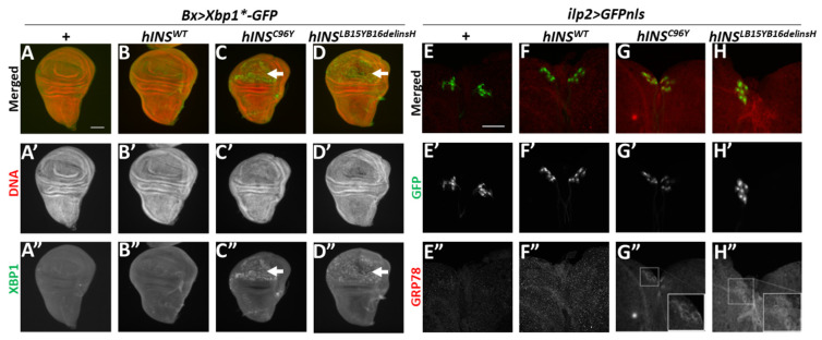
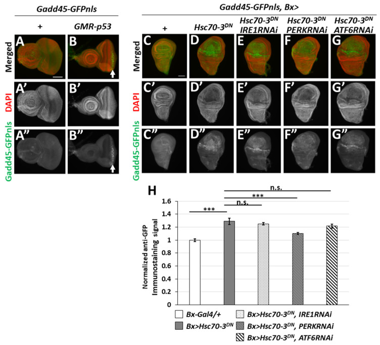
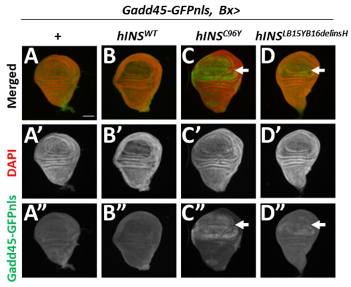
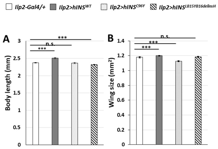
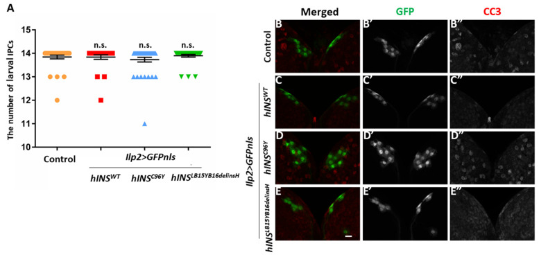
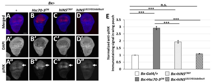
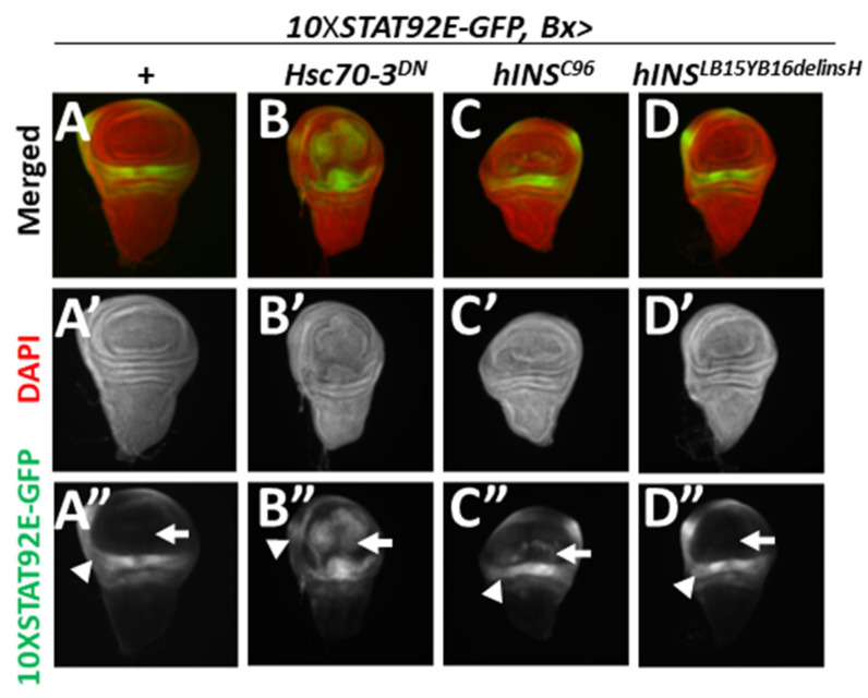
Mutations in the insulin gene (INS) are frequently associated with human permanent neonatal diabetes mellitus. However, the mechanisms underlying the onset of this genetic disease is not sufficiently decoded. We induced expression of two types of human mutant INSs in Drosophila using its ectopic expression system and investigated the resultant responses in development. Expression of the wild-type preproinsulin in the insulin-producing cells (IPCs) throughout the larval stage led to a stimulation of the overall and wing growth. However, ectopic expression of human mutant preproinsulins, hINSC96Y and hINSLB15YB16delinsH, neither of which secreted from the β-cells, could not stimulate the Drosophila growth. Furthermore, neither of the mutant polypeptides induced caspase activation leading to apoptosis. Instead, they induced expression of several markers indicating the activation of unfolded protein response, such as ER stress-dependent Xbp1 mRNA splicing and ER chaperone induction. We newly found that the mutant polypeptides induced the expression of Growth arrest and DNA-damage-inducible 45 (Gadd45) in imaginal disc cells. ER stress induced by hINSC96Y also activated the JAK-STAT signaling, involved in inflammatory responses. Collectively, we speculate that the diabetes-like growth defects appeared as a consequence of the human mutant preproinsulin expression was involved in dysfunction of the IPCs, rather than apoptosis.

Keywords: Drosophila; ER stress; Gadd45; JNK; NDM; diabetes.


Conflict of interest statement

The authors declare no competing interests.

Figures

Figure 1
Figure 1
Induction of UPR observed in wing imaginal discs and larval IPCs harboring the ectopic expressing human mutant preproinsulin. (AD) Xbp1-GFP fluorescence (green in (AD), white in (A”D”), arrows in (C,C”) and (D,D”)) produced because of ER stress-dependent splicing of Xbp1*-GFP mRNA in wing imaginal discs. Red in (AD) (white in (A’D’)): DNA staining. (AA”) A control wing imaginal disc (Bx-Gal4/+; UAS-Xbp1*-GFP). (BB”). A wing imaginal disc expressing hINSWT in the wing pouch region (Bx > hINSWT, Xbp1*-GFP). (CC”) A wing imaginal disc expressing hINSC96Y (Bx > hINSC96Y, Xbp1*-GFP). (DD”) A wing imaginal disc expressing hINSLB15YB16delinsH (Bx > hINSLB15YB16delinsH, Xbp1*-GFP). Scale bar: 100 μm. (EH) Anti-GRP78 immunostaining (red in (EH), white in (E”H”)) of brains the ectopically expressing GFPnls in IPCs from third-instar larvae. IPC nuclei are visualized by GFP expression (green in (EH), white in (E’H’)). (EE”) A control larval brain (ilp2 > GFPnls). (FF”) A Larval brain with IPC-specific expression of hINSWT (ilp2 > hINSWT, GFPnls). (GG”) A Larval brain expressing hINSC96Y in IPCs (ilp2 > hINSC96Y, GFPnls). (HH”) A Larval brain expressing hINSLB15YB16delinsH in IPCs (ilp2 > hINSLB15YB16delinsH, GFPnls). Insets in (G”) and (H”) present a magnified view of the IPC cluster. Scale bar: 50 μm.
Figure 2
Figure 2
p53-Dependent Gadd45 expression and a requirement of three ER stress sensors for ER-stress induced Gadd45 expression in wing imaginal discs. (A,B) Induction of Gadd45-GFP (green in (A,B), white in (A”,B”), arrow in (B,B”)) generated by p53-dependent cellular stress response. Red in (A,B) (white in (A’,B’)): DNA staining. (AA”) A control eye imaginal disc (Gadd45-GFP). (BB”) An eye imaginal disc expressing p53 in after a morphogenic furrow of eye imaginal discs under the GMR promoter. Scale bar: 100 μm (CG). Fluorescent micrographs of wing imaginal discs harboring Gadd45-GFP reporter. DAPI staining (red). (CC”) A Control wing disc (Bx-Gal4/+; Gadd45-GFP/+). (DD”) A wing disc having Hsc70-3DN expression in the wing pouch region (Gadd45-GFP, Bx > Hsc70-3DN). (EE”) A wing disc having Hsc70-3DN expression and IRE1 depletion (Gadd45-GFP, Bx > Hsc70-3DN, IRE1RNAi). (FF”) A wing disc having Hsc70-3DN expression and PERK depletion (Gadd45-GFP, Bx > Hsc70-3DN, PERKRNAi). (GG”) A wing disc having Hsc70-3DN expression, ATF6 depletion (Gadd45-GFP, Bx > Hsc70-3DN, ATF6RNAi). Scale bar: 100 μm. (H) Quantification of GFP levels in wing discs. The intensity of Gadd45-GFP fluorescence in each wing imaginal disc expressing Hsc70-3DN and/or dsRNA against each of three ER stress sensors as calculated and normalized to that of the control, set to 1.0 (Bx-Gal4/+) (n > 21, *** p < 0.001, n.s.: not significant, Student’s t-test). Error bars represent SEMs.
Figure 3
Figure 3
The ectopic expression of human mutant preproinsulin, hINSC96Y, and hINSLB15YB16delinsH induced Gadd45 transcription in wing imaginal discs. (A,B) GFP fluorescence (green in (A,B), white in (A”D”), arrow in (C,C”) and (D,D”)) indicating Gadd45 expression in wing imaginal discs. Red in AD: DNA staining (white in A’D’). (AA”) A control wing disc (Bx-Gal4/+; Gadd45-GFP/+). (BB”) A wing disc expressing hINSWT in the wing pouch region (Gadd45-GFP, Bx > hINSWT). (CC”) A wing disc expressing hINSC96Y (Gadd45-GFP, Bx > hINSC96Y). (DD”) A wing disc expressing hINSLB15YB16delinsH (Gadd45-GFP, Bx > hINSLB15YB16delinsH). Scale bar: 100 μm.
Figure 4
Figure 4
The ectopic expression of human mutant preproinsulin in IPCs resulted in the production of smaller adults indicating growth inhibition. (A,B) Adult phenotypes in male flies derived from individuals harboring targeted expression of human preproinsulin in IPCs throughout development. Note that the expression of hINSWT in IPCs stimulated Drosophila growth, while either of hINSC96Y or hINSLB15YB16delinsH failed. (A) Quantification of body length of male adults (n > 69, n.s.: not significant, *** p < 0.001, Student’s t-test). Error bars represent SEMs. (B) Quantification of wing size of the male adults (n > 113, n.s.: not significant, *** p < 0.001, Student’s t-test). Error bars represent SEMs.
Figure 5
Figure 5
The ectopic expression of neither of hINSC96Y or hINSLB15YB16delinsH induced caspase activation or the loss of larval IPCs. (A) The number of larval IPCs in control larvae and larvae expressing human preproinsulin at the third-instar stage. Note that the differences in the IPC number between larvae expressing every three types of preproinsulin and control larvae (ilp2 > GFPnls) were not significant (n > 25, n.s.: not significant, Mann–Whitney’s U-test). (BE) Immunostaining of third-instar larval brains having IPCs labeled with GFPnls with anti-Cleaved Caspase-3 (CC3) antibody. Red in B–E (white in (B”E”)): Anti-CC3 immunostaining signal. Green I n (BE) (white in (B’E’)): IPCs visualized by expression of GFPnls. (BB”) Control larval brain (ilp2 > GFPnls). (CC”) Larval brain the ectopic expressing hINSWT in IPCs (ilp2 > hINSWT, GFPnls). (DD”) Larval brain with IPCs-specific expression of hINSC96Y (ilp2 > hINSC96Y, GFPnls). (EE”) Larval brain with IPCs-specific expression of hINSLB15YB16delinsH (ilp2 > hINSLB15YB16delinsH, GFPnls). Scale bar: 10 μm.
Figure 6
Figure 6
The ectopic expression of hINSC96Y can activate JNK pathway in wing imaginal discs. (AD) Anti-pJNK immunostaining of wing imaginal discs having an accumulation of ER stress in third-instar larvae. (AA”) A control wing disc (Bx-Gal4/+). (BB”) A wing discs expressing Hsc70-3DN (Bx > Hsc70-3DN). (CC”) A wing disc expressing hINSC96Y (Bx > hINSC96Y). (DD”) A wing disc expressing hINSLB15YB16delinsH (Bx > hINSLB15YB16delinsH). Anti-pJNK immunostaining signal and DNA staining are colored in red (white in (A”D”)) and blue (white in (A’D’)), respectively. Scale bar: 100 μm. (E) Quantification of pJNK signals in wing imaginal discs. The intensity of anti-pJNK immunofluorescence was calculated and normalized to the control value, which was set as 1.0 (Bx-Gal4/+) (n > 15, *** p < 0.001, n.s.: not significant, Student’s t-test) Error bars represent SEMs.
Figure 7
Figure 7
Activation of JAK/STAT signaling pathway observed in wing imaginal discs with the ectopic expression of Hsc70-3DN or hINSC96Y. (AD) GFP fluorescence (Green in (AD), white in (AB”)) indicating activation of JAK/STAT signaling pathway in wing imaginal discs. Red in (AD) (white in (A’D’)): DNA staining. (AA”) A control wing imaginal disc (Bx-Gal4/+; 10XSTAT92E-GFP). (BB”) A wing disc expressing Hsc70-3DN (10XSTAT92E-GFP, Bx > Hsc70-3DN). (CC”) A wing disc expressing hINSC96Y (10XSTAT92E-GFP, Bx > hINSC96Y). (DD”) A wing disc expressing hINSLB15YB16delinsH (10XSTAT92E-GFP, Bx > hINSLB15YB16delinsH).

References

    1. Collares C.V., Evangelista A.F., Xavier D.J., Rassi D.M., Arns T., Foss-Freitas M.C., Foss M.C., Puthier D., Sakamoto-Hojo E.T., A Passos G., et al. Identifying common and specific microRNAs expressed in peripheral blood mononuclear cell of type 1, type 2, and gestational diabetes mellitus patients. BMC Res. Notes. 2013;6:491. doi: 10.1186/1756-0500-6-491. - DOI - PMC - PubMed
    1. Cerf M.E. Beta Cell Dysfunction and Insulin Resistance. Front. Endocrinol. 2013;4:37. doi: 10.3389/fendo.2013.00037. - DOI - PMC - PubMed
    1. Colombo C., Porzio O., Liu M., Massa O., Vasta M., Salardi S., Beccaria L., Monciotti C., Toni S., Pedersen O., et al. Seven mutations in the human insulin gene linked to permanent neonatal/infancy-onset diabetes mellitus. J. Clin. Investig. 2008;118:2148–2156. doi: 10.1172/JCI33777. - DOI - PMC - PubMed
    1. Støy J., Edghill E.L., Flanagan S.E., Ye H., Paz V.P., Pluzhnikov A., Below J.E., Hayes M.G., Cox N.J., Lipkind G.M., et al. Insulin gene mutations as a cause of permanent neonatal diabetes. Proc. Natl. Acad. Sci. USA. 2007;104:15040–15044. doi: 10.1073/pnas.0707291104. - DOI - PMC - PubMed
    1. Støy J., Steiner D.F., Park S.-Y., Ye H., Philipson L.H., Bell G.I. Clinical and molecular genetics of neonatal diabetes due to mutations in the insulin gene. Rev. Endocr. Metab. Disord. 2010;11:205–215. doi: 10.1007/s11154-010-9151-3. - DOI - PMC - PubMed

MeSH terms