

Impact of Lung Segmentation on the Diagnosis and Explanation of COVID-19 in Chest X-ray Images
- PMID: 34770423
- PMCID: PMC8587284
- DOI: 10.3390/s21217116
Impact of Lung Segmentation on the Diagnosis and Explanation of COVID-19 in Chest X-ray Images
Abstract
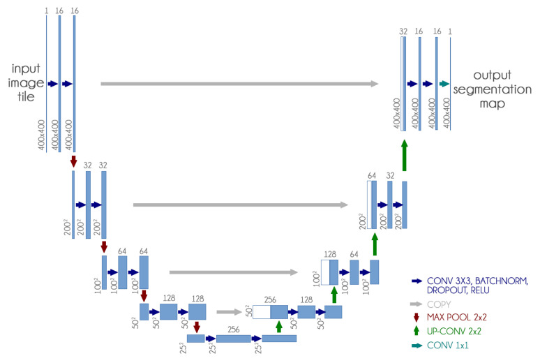
COVID-19 frequently provokes pneumonia, which can be diagnosed using imaging exams. Chest X-ray (CXR) is often useful because it is cheap, fast, widespread, and uses less radiation. Here, we demonstrate the impact of lung segmentation in COVID-19 identification using CXR images and evaluate which contents of the image influenced the most. Semantic segmentation was performed using a U-Net CNN architecture, and the classification using three CNN architectures (VGG, ResNet, and Inception). Explainable Artificial Intelligence techniques were employed to estimate the impact of segmentation. A three-classes database was composed: lung opacity (pneumonia), COVID-19, and normal. We assessed the impact of creating a CXR image database from different sources, and the COVID-19 generalization from one source to another. The segmentation achieved a Jaccard distance of 0.034 and a Dice coefficient of 0.982. The classification using segmented images achieved an F1-Score of 0.88 for the multi-class setup, and 0.83 for COVID-19 identification. In the cross-dataset scenario, we obtained an F1-Score of 0.74 and an area under the ROC curve of 0.9 for COVID-19 identification using segmented images. Experiments support the conclusion that even after segmentation, there is a strong bias introduced by underlying factors from different sources.
Keywords: COVID-19; chest X-ray; explainable artificial intelligence; semantic segmentation.
Conflict of interest statement
The authors declare no conflict of interest.
Figures


References
-
- Lauer S.A., Grantz K.H., Bi Q., Jones F.K., Zheng Q., Meredith H.R., Azman A.S., Reich N.G., Lessler J. The incubation period of coronavirus disease 2019 (COVID-19) from publicly reported confirmed cases: Estimation and application. Ann. Intern. Med. 2020;172:577–582. doi: 10.7326/M20-0504. - DOI - PMC - PubMed
-
- Self W.H., Courtney D.M., McNaughton C.D., Wunderink R.G., Kline J.A. High discordance of chest X-ray and computed tomography for detection of pulmonary opacities in ED patients: Implications for diagnosing pneumonia. Am. J. Emerg. Med. 2013;31:401–405. doi: 10.1016/j.ajem.2012.08.041. - DOI - PMC - PubMed
MeSH terms
Grants and funding
LinkOut - more resources
Full Text Sources
Medical
