. 2021 Nov 3;26(21):6668.
doi: 10.3390/molecules26216668.

A New Class of Uracil-DNA Glycosylase Inhibitors Active against Human and Vaccinia Virus Enzyme

Affiliations

A New Class of Uracil-DNA Glycosylase Inhibitors Active against Human and Vaccinia Virus Enzyme

Inga R Grin et al. Molecules. .

Abstract

Uracil-DNA glycosylases are enzymes that excise uracil bases appearing in DNA as a result of cytosine deamination or accidental dUMP incorporation from the dUTP pool. The activity of Family 1 uracil-DNA glycosylase (UNG) activity limits the efficiency of antimetabolite drugs and is essential for virulence in some bacterial and viral infections. Thus, UNG is regarded as a promising target for antitumor, antiviral, antibacterial, and antiprotozoal drugs. Most UNG inhibitors presently developed are based on the uracil base linked to various substituents, yet new pharmacophores are wanted to target a wide range of UNGs. We have conducted virtual screening of a 1,027,767-ligand library and biochemically screened the best hits for the inhibitory activity against human and vaccinia virus UNG enzymes. Although even the best inhibitors had IC50 ≥ 100 μM, they were highly enriched in a common fragment, tetrahydro-2,4,6-trioxopyrimidinylidene (PyO3). In silico, PyO3 preferably docked into the enzyme's active site, and in kinetic experiments, the inhibition was better consistent with the competitive mechanism. The toxicity of two best inhibitors for human cells was independent of the presence of methotrexate, which is consistent with the hypothesis that dUMP in genomic DNA is less toxic for the cell than strand breaks arising from the massive removal of uracil. We conclude that PyO3 may be a novel pharmacophore with the potential for development into UNG-targeting agents.

Keywords: DNA repair; inhibitors; pyrimidines; uracil–DNA glycosylase; virtual screening.


Conflict of interest statement

The authors declare no conflict of interest. The funders had no role in the design of the study; in the collection, analyses, or interpretation of data; in the writing of the manuscript, or in the decision to publish the results.

Figures
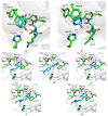
Figure 6
Figure 6
Structures of the uracil-binding pockets of (a) hUNG (Protein Data Bank ID 1SSP [44]) and (b) vvUNG (4YIG [47]) with the excised Ura base. Both structures also contain the abasic DNA, which is not shown for clarity. (cg) Structure of vvUNG uracil-binding pocket with docked compounds 2D (c), 3A (d), 3B (e), 4B (f), and 4F (g). Dashes indicate hydrogen bonds; their length in Å is shown. The carbon atoms of the excised uracil base (ab) and the inhibitors (cg) are colored cyan; protein carbons are green.
Figure 1
Figure 1
Docking of inhibitors into hUNG structure. (a) Illustration of the On-Top Docking procedure: the ΔG-score calculated by Lead Finder for each of the selected compounds for a set of poses inside the binding site (cyan bars) and on the protein surface (red bars); * marks the potentially inactive compounds, for which pMedian Test > 0.01 (see Table S1). (b) Structure of hUNG (PDB ID 3FCF [29]) with compound 4B docked into the active site. Lengths of the stabilizing hydrogen bonds are shown. Double-headed arrows indicate interactions with the π-system of the PyO3 moiety. (c) Same as (b) but with compound 4F docked into the hUNG active site.
Figure 2
Figure 2
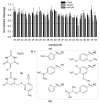
Biochemical screening of the inhibitors identified by molecular docking. (a) hUNG activity in the presence of the inhibitors at 10 μM (black bars), 100 μM (light gray bars), or 1000 μM (dark gray bars). The activity is normalized for that in the absence of the inhibitor. Means and SD of three independent experiments are shown; (b) Structures of the compounds 2D, 3A, 3B, 4B, 4E, 4F, and 4G.
Figure 3
Figure 3
The mode of UNG inhibition. (a) Cleavage of the substrate (1 μM) by hUNG at different concentrations of compound 4B; (b) cleavage by hUNG at different concentrations of the substrate and compound 4B plotted in double reciprocal coordinates.
Figure 4
Figure 4
Cytotoxicity of UNG inhibitors. (a) Survival of HEK 293FT cells in the presence of the UNG inhibitors 4B and 4F (0.8–500 μM); (b) survival of HEK 293FT cells in the presence of MTX alone (0.02–10 μM) or 100 μM 4B plus MTX (0.1–10 μM); (c) survival of HEK 293FT cells in the presence of the UNG inhibitors 4B and 4F (0.8–500 μM) plus 0.1 μM MTX. In Panel C, the survival in the presence of 0.1 μM MTX alone is taken for 100%. Mean ± s. e. m. is shown (n = 3).
Figure 5
Figure 5
Biochemical screening of the inhibitors against vvUNG. The inhibitor concentration was 10 μM (black bars), 100 μM (light gray bars), or 1000 μM (dark gray bars). The activity is normalized for that in the absence of the inhibitor. Means and SD of three independent experiments are shown.

References

    1. Aravind L., Koonin E.V. The α/β fold uracil DNA glycosylases: A common origin with diverse fates. Genome Biol. 2000;1:research0007. doi: 10.1186/gb-2000-1-4-research0007. - DOI - PMC - PubMed
    1. Visnes T., Doseth B., Pettersen H.S., Hagen L., Sousa M.M.L., Akbari M., Otterlei M., Kavli B., Slupphaug G., Krokan H.E. Uracil in DNA and its processing by different DNA glycosylases. Philos. Trans. R. Soc. Lond. B Biol. Sci. 2009;364:563–568. doi: 10.1098/rstb.2008.0186. - DOI - PMC - PubMed
    1. Kavli B., Otterlei M., Slupphaug G., Krokan H.E. Uracil in DNA—General mutagen, but normal intermediate in acquired immunity. DNA Repair. 2007;6:505–516. doi: 10.1016/j.dnarep.2006.10.014. - DOI - PubMed
    1. Friedberg E.C., Walker G.C., Siede W., Wood R.D., Schultz R.A., Ellenberger T. DNA Repair and Mutagenesis. ASM Press; Washington, DC, USA: 2006. p. 1118.
    1. Zharkov D.O. Base excision DNA repair. Cell. Mol. Life Sci. 2008;65:1544–1565. doi: 10.1007/s00018-008-7543-2. - DOI - PMC - PubMed

LinkOut - more resources