. 2021 Oct 22;13(21):5308.
doi: 10.3390/cancers13215308.

New Therapeutic Strategy for Overcoming Multidrug Resistance in Cancer Cells with Pyrazolo[3,4- d]pyrimidine Tyrosine Kinase Inhibitors

Affiliations

New Therapeutic Strategy for Overcoming Multidrug Resistance in Cancer Cells with Pyrazolo[3,4- d]pyrimidine Tyrosine Kinase Inhibitors

Ana Podolski-Renić et al. Cancers (Basel). .

Abstract

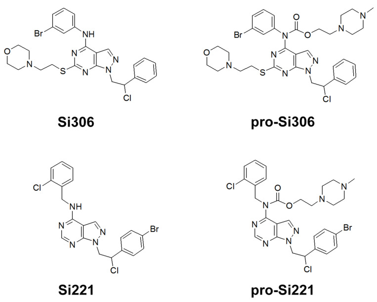
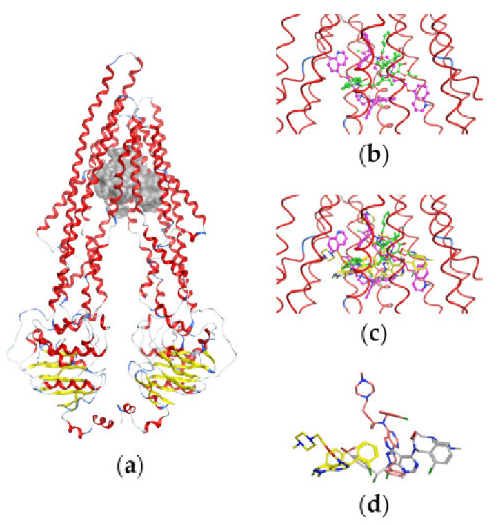
Tyrosine kinase inhibitors (TKIs) often interact with the multidrug resistant (MDR) phenotype of cancer cells. In some cases, TKIs increase the susceptibility of MDR cancer cells to chemotherapy. As the overexpression of membrane transporter P-glycoprotein (P-gp) is the most common alteration in MDR cancer cells, we investigated the effects of TKI pyrazolo[3,4-d]pyrimidines on P-gp inhibition in two cellular models comprising sensitive and corresponding MDR cancer cells (human non-small cell lung carcinoma and colorectal adenocarcinoma). Tested TKIs showed collateral sensitivity by inducing stronger inhibition of MDR cancer cell line viability. Moreover, TKIs directly interacted with P-gp and inhibited its ATPase activity. Their potential P-gp binding site was proposed by molecular docking simulations. TKIs reversed resistance to doxorubicin and paclitaxel in a concentration-dependent manner. The expression studies excluded the indirect effect of TKIs on P-gp through regulation of its expression. A kinetics study showed that TKIs decreased P-gp activity and this effect was sustained for seven days in both MDR models. Therefore, pyrazolo[3,4-d]pyrimidines with potential for reversing P-gp-mediated MDR even in prolonged treatments can be considered a new therapeutic strategy for overcoming cancer MDR.

Keywords: P-glycoprotein inhibitors; Src family tyrosine kinase inhibitors; cancer; multidrug resistance.


Conflict of interest statement

The authors declare no conflict of interest.

Figures

Figure 1
Figure 1
Chemical structures of Si306, pro-Si306, Si221, and pro-Si221.
Figure 2
Figure 2
SFK inhibitors suppress P-gp activity. Flow-cytometric profile and the mean fluorescence intensity of Rho123 accumulation assessed after 30-min treatment with 10 µM Si306, pro-Si306, Si221, and pro-Si221 in (a) NCI-H460/R and (b) DLD1-TxR cell lines. Dasatinib and Dex-VER are included as reference compounds. Three independent experiments were performed, and a minimum of 10,000 events were collected for each experimental sample. The fluorescence intensity of Rho123 accumulation is presented as the mean ± SEM. Statistically significant differences between untreated control and treated samples: p < 0.01 (**), p < 0.001 (***).
Figure 3
Figure 3
Concentration-dependent inhibition of P-gp activity. Rho 123 accumulation in (a) NCI-H460/R and (b) DLD1-TxR cell lines was assessed after the application of increasing concentrations of Si306, pro-Si306, and pro-Si221 (1, 2, 5, 10, 20 µM). The lower range of concentrations for (c) pro-Si221 (0.05, 0.1, 0.2, 0.5, 1 μM) and (d) TQ (1, 2, 5, 10, 20 nM) in NCI-H460/R and DLD1-TxR cells. The percentage of P-gp inhibition is expressed as an increase in Rho 123 accumulation; 100% of P-gp inhibition is considered when the level of Rho123 accumulation achieves the level of Rho123 accumulation in sensitive cells. Three independent experiments were performed (a minimum of 10,000 events were collected for each experimental sample). A statistically significant difference between 5 µM and 20 µM treatments with Si306, pro-Si306, and pro-Si221 is presented in (a) and (b): p < 0.01 (**), p < 0.001 (***), not significant (n.s.).
Figure 4
Figure 4
SFK inhibitors affect the ATPase activity of P-gp. The samples were treated with 5 µM and 20 µM Si306, pro-Si306, and pro-Si221. Verapamil was used as a positive control. The y-axis represents the difference in relative light units (ΔRLU) between Na3VO4-treated samples and samples treated with the tested compounds. ΔRLU basal is the difference in the luminescent signal between Na3VO4-treated and untreated samples. The results are expressed as the mean ± SEM. Statistically significant differences between 5 µM and 20 µM treatments with Si306, pro-Si306, and pro-Si221: p < 0.05 (*), not significant (n.s.).
Figure 5
Figure 5
The effect of SFK inhibitors on P-gp expression at the protein level. Mean fluorescence intensity of primary P-gp antibody in NCI-H460/R and DLD1-TxR cells assessed after 48-h treatment with 5 µM and 10 µM (treatment 1 and treatment 2) for Si306 and pro-Si306 as well as 1 µM and 2 µM (treatment 1 and treatment 2) for pro-Si221. TQ is included as a reference compound with 5 nM for treatment 1 and 10 nM for treatment 2. The negative control is the auto-fluorescence of unstained untreated cells, whereas the positive control represents the fluorescence of anti-P-gp-labeled untreated cells. The results are expressed as the mean ± SEM. Statistically significant differences between the untreated control and treated samples calculated from three independent experiments: p < 0.001 (***).
Figure 6
Figure 6
Binding of pro-Si221 in the P-gp cavity. (a) A front view of the P-gp structure used in docking studies with the bound substrate PTX (in green) and two molecules of the inhibitor Zosuquidar (in magenta); the protein backbone is shown as a ribbon and is colored according to the secondary structure—loop (in grey), helix (in red), and turn (in blue). (b) A closer view of the transmembrane-binding cavity of P-gp with bound PTX and two molecules of Zosuquidar (shown as balls and sticks). (c) A closer view of the transmembrane-binding cavity of P-gp with bound PTX, Zosuquidar, and three conformations of pro-Si221 (yellow sticks) corresponding to three different binding poses among the 10 best generated during docking. (d) The three different poses of pro-Si221 (shown as yellow, orange, and light gray sticks) positioned equally to those on (c). For a better view, the protein backbone is shown as a line ribbon in (b,c).
Figure 7
Figure 7
Sustained inhibition of P-gp after acute treatments with pro-Si306 and pro-Si221. Mean fluorescence intensity of Rho 123 accumulation assessed in NCI-H460/R and DLD1-TxR cells treated with pro-Si306 and pro-Si221 for 24 h, 48 h, 96 h, and 168 h. TQ was used as a reference compound. Three independent experiments were performed (a minimum of 10,000 events were collected for each experimental sample).

References

    1. Robey R.W., Pluchino K.M., Hall M.D., Fojo A.T., Bates S.E., Gottesman M.M. Revisiting the role of ABC transporters in multidrug-resistant cancer. Nat. Rev. Cancer. 2018;18:452–464. doi: 10.1038/s41568-018-0005-8. - DOI - PMC - PubMed
    1. Fletcher J.I., Williams R.T., Henderson M.J., Norris M.D., Haber M. ABC transporters as mediators of drug resistance and contributors to cancer cell biology. Drug Resist. Updates Rev. Comment. Antimicrob. Anticancer Chemother. 2016;26:1–9. doi: 10.1016/j.drup.2016.03.001. - DOI - PubMed
    1. Ogihara T., Mizoi K., Kamioka H., Yano K. Physiological Roles of ERM Proteins and Transcriptional Regulators in Supporting Membrane Expression of Efflux Transporters as Factors of Drug Resistance in Cancer. Cancers. 2020;12:3352. doi: 10.3390/cancers12113352. - DOI - PMC - PubMed
    1. Li W., Zhang H., Assaraf Y.G., Zhao K., Xu X., Xie J., Yang D.H., Chen Z.S. Overcoming ABC transporter-mediated multidrug resistance: Molecular mechanisms and novel therapeutic drug strategies. Drug Resist. Updates Rev. Comment. Antimicrob. Anticancer Chemother. 2016;27:14–29. doi: 10.1016/j.drup.2016.05.001. - DOI - PubMed
    1. Kodan A., Yamaguchi T., Nakatsu T., Matsuoka K., Kimura Y., Ueda K., Kato H. Inward- and outward-facing X-ray crystal structures of homodimeric P-glycoprotein CmABCB1. Nat. Commun. 2019;10:88. doi: 10.1038/s41467-018-08007-x. - DOI - PMC - PubMed