. 2021 Oct 24;13(21):5338.
doi: 10.3390/cancers13215338.

IL-17B/RB Activation in Pancreatic Stellate Cells Promotes Pancreatic Cancer Metabolism and Growth

Affiliations

IL-17B/RB Activation in Pancreatic Stellate Cells Promotes Pancreatic Cancer Metabolism and Growth

Jiahui Li et al. Cancers (Basel). .

Abstract

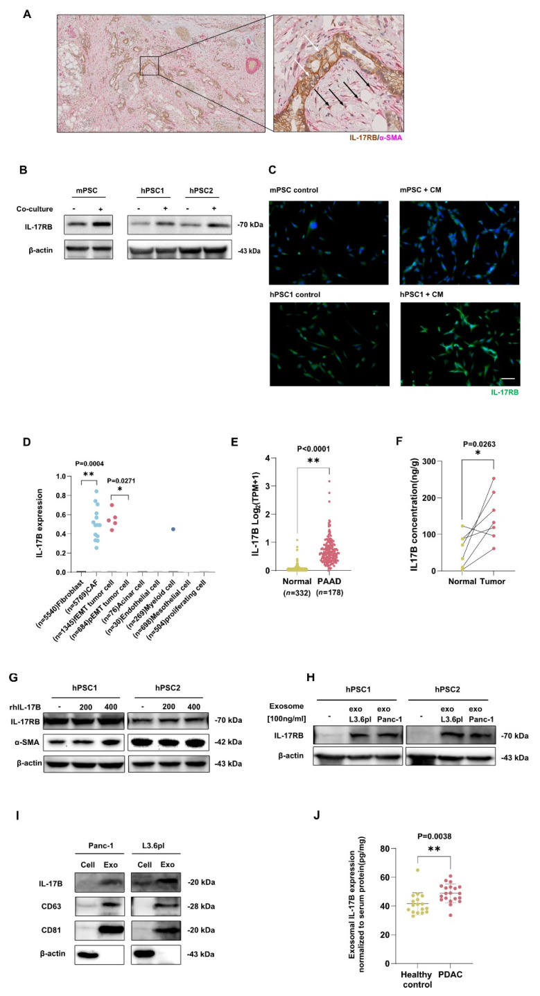
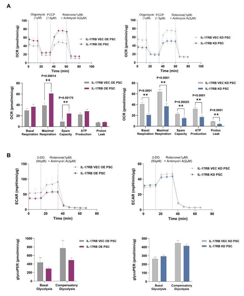
In pancreatic ductal adenocarcinoma (PDAC), the tumor stroma constitutes most of the cell mass and contributes to therapy resistance and progression. Here we show a hitherto unknown metabolic cooperation between pancreatic stellate cells (PSCs) and tumor cells through Interleukin 17B/Interleukin 17B receptor (IL-17B/IL-17RB) signaling. Tumor-derived IL-17B carrying extracellular vesicles (EVs) activated stromal PSCs and induced the expression of IL-17RB. PSCs increased oxidative phosphorylation while reducing mitochondrial turnover. PSCs activated tumor cells in a feedback loop. Tumor cells subsequently increased oxidative phosphorylation and decreased glycolysis partially via IL-6. In vivo, IL-17RB overexpression in PSCs accelerated tumor growth in a co-injection xenograft mouse model. Our results demonstrate a tumor-to-stroma feedback loop increasing tumor metabolism to accelerate tumor growth under optimal nutritional conditions.

Keywords: IL17B/RB; metabolism; pancreatic cancer; tumor microenvironment.


Conflict of interest statement

None declared. The authors declare no competing interest.

Figures

Figure 1
Figure 1
Expression of IL-17RB in PDAC tumors and stroma from humans and mice. (A) Double-immunohistolochemistry staining of IL-17RB (brown) and α-SMA. White arrows point to IL-17RB positive tumor cells. Black arrows depict double-positive stromal cells. Scale bars = 100 μm; (B) Murine PSCs express more IL-17RB after co-culture with Panc02 pancreatic cancer cells than naive PSCs (left). Two human primary PSCs preparations (hPSC1 and hPSC2) express more IL-17RB after co-culture with L3.6pl pancreatic cancer cells than naive PSCs (right). Western blots of mouse PSCs (RLT-PSCs) and two primary human PSC preparations from surgical specimens. Data show typical images from three independent experiments; (C) PSCs increase IL-17RB expression when treated with conditioned medium (CM) from tumor cells. IL-17RB immunofluorescence staining (green) of naive murine PSCs compared to PSCs treated with CM from Panc02 tumor cells for 48 h (upper row). A human primary PSC preparation increases IL-17RB expression when cultured for 48 h with conditioned CM from L3.6pl tumor cells (lower row). Scale bar = 100 μm; (D) Stromal cells express IL-17B. IL-17B expression by cell type in human PDAC on single-cell RNA sequencing of a public database (Genome Sequence Archive PRJCA001063). Each dot represents a single cell expressing IL-17B. PSCs are contained in the fibroblast compartment in healthy tissue and differentiate into CAFs in the tumor environment. In the first two columns, fibroblasts from healthy tissue were compared with CAFs. Fibroblast (n = 5540) vs. CAF (n = 5769; p = 0.004 t-test), tumor cells in full epithelial-to-mesenchymal transition (fEMT, n = 1345) vs. tumor cells in partial epithelial-to-mesenchymal transition (pEMT, n = 684; p = 0.0271, t-test); (E) IL-17B expression in tumor and paired non-malignant pancreatic tissues from publicly available TCGA and GTEx data sets analyzed with the GEPIA2 portal; (F) Increased IL-17B expression in pancreatic cancer tissue compared to adjacent non-tumor tissue (normal) tissue. ELISA of specimens of in-house PDAC patients (n = 7; p = 0.0263; Wilcoxon matched-pairs test); (G) Western blot on human primary PSC preparations of IL-17RB and α-SMA after stimulation with 200 respective 400 ng/mL rhIL-17B; (H) Extracellular vesicles (EVs) from human pancreatic cancer cell lines induced the expression of IL-17RB in PSCs. IL-17RB Western blot of PSCs treated with EVs isolated from two human pancreatic cancer cell lines (L3.6pl and Panc-1). The Data represent the typical images of three independent experiments; (I) CD63 and CD81 are specific markers for extracellular vesicles. Data represent the typical images of three independent experiments; (J) ELISA of exosome IL-17B expression normalized to serum. * p < 0.05, ** p < 0.01.
Figure 2
Figure 2
IL-17RB alters mitochondrial turnover and IL-6 secretion. (A) IL-17RB OE PSCs stably express IL-17RB without activation. IL-17RB KD PSCs do not express IL-17RB; (B) IL-17RB OE PSCs increase α-SMA. IL-17RB and α-SMA Western blot of IL-17RB OE and IL-17KD cells with corresponding control of IL-17RB VEC OE and IL-17RB VEC KD cells; (C) IL-17RB overexpression increases PSCs proliferation. MTT assay comparing IL-17RB OE, IL-17RB KD PSCs with respective controls (IL-17RB VEC OE and VEC KD, p < 0.0001, Mann–Whitney U test). Data represent the mean of three independent experiments conducted in sextuplicate; (D) IL-17RB overexpression decreases IL-6 expression. IL-6 Western blot of IL-17RB OE and IL-17KD PSCs with corresponding controls; (E) IL-17RB overexpression decreases IL-6 secretion, while IL-17RB knockdown induces no changes. ELISA of cell culture supernatant (n = 3, OE vs. VEC OE, p = 0.0167, Mann-Whitney U test); (F) Auto-/mitophagy Western blot of IL-17RB OE and IL-17KD PSCs with corresponding controls. p62—Autophagy receptor p62, PRK8—Ubiquitin E3 ligase Parkin; (G) IL-17RB overexpression reduces mitochondrial fission and fragmentation. Western blot of IL-17RB OE and IL-17RB KD PSCs with corresponding controls. pMMF—phospho-mitochondrial fission factor, DRP1—phospho-Dynamin related protein 1; (H) IL-17RB overexpression induces elongated mitochondria with tubular cristae (white arrows). Electron microscope images of IL-17RB OE PSCs compared to IL-17RB VEC OE controls. Scale bar = 250 nm. * p < 0.05, ** p < 0.01.
Figure 3
Figure 3
IL-17RB overexpression induces mitochondrial OXPHOS in PSCs (A) IL-17RB overexpression increases and IL-17RB knockdown decreases mitochondrial respiration in PSCs. Oxygen consumption rate (OCR) was measured with the Seahorse mitostress test. Seahorse profiles (upper panel) and statistical analysis (unpaired t-test, lower panel). Data represent the mean (±SD) of three independent experiments conducted in quintuplicate; (B) IL-17RB overexpression decreases glycolysis in PSCs while IL-17RB knockdown induces no changes. Statistical significance was not reached in either group. The glycolytic proton efflux rate (glycoPER) correlates with lactate accumulation over time, showing glycolysis: seahorse profiles (upper panel) and statistical analysis (unpaired t-test, lower panel). Data represent the mean (±SD) of three independent experiments conducted in quintuplicate. ** p < 0.01.
Figure 4
Figure 4
IL-17RB expressing PSCs promote tumor growth (A) Immunohistochemistry staining of pan-cytokeratin (brown, upper panel) and hematoxylin and eosin (H&E) of tumors developing after co-injection of PSCs and Mia PaCa-2 cells; (B) Control IL-17RB VEC OE PSCs and IL-17RB OE PSCs accelerate the growth of MIA PaCa-2 tumors. Image of tumors harvested at day 46 from NSG mice after co-injecting MIA PaCa-2 cells and IL-17RB OE PSCs (OE, n = 8) respective control PSCs (VEC OE, n = 8, upper panel). Statistics of tumor volume (n = 8; Mann-Whitney U test, p = 0.0024 MIA PaCa-2 vs. VEC OE, p = 0.0065 VEC OE vs. OE, lower left panel) and weight (n = 8; Mann-Whitney U test, p = 0.0045 MIA PaCa-2 vs. VEC OE, p = 0.0099 VEC OE vs. OE) at day 46 after injection (lower right panel). Scale bars = 1 cm; (C) Tumor growth curve of animals injected with MIA PaCa2-cells or tumor cells with OE PSCs or VEC OE PSCs (all groups, n = 8). VEC OE PSC and OE PSC curve significantly diverged from MIA PaCa-2 tumors at day 10, comparing individual time points with t-tests. The OE PSC curve significantly diverged from the VEC OE PSC curve on day 26; (D) The conditioned medium (CM) of IL-17RB overexpressing PSCs (CM OE) increases the growth of L3.6pl pancreatic cancer cells. CM of control PSCs transfected with the corresponding empty vector (CM VEC OE) did not. The conditioned medium of IL-17RB knockdown PSCs (CM KD) increases tumor cell growth to the same extent as corresponding CM from control PSCs (CM VEC KD). MTT cell assay to assess cell proliferation; (E) Conditioned medium from IL-17RB OE PSCs but not from IL-17RB KD PSCs increases CXCR4 expression in L3.6pl pancreatic cancer cells. Flow cytometry analysis of FSC-A/CXCR4 positive cells. Data represent the mean (±SD) of three independent experiments performed in triplicate; (F) IL-17RB overexpression inhibits the IL-6 signaling pathway. Western blot of MIAPaCa-2 cells treated with the conditioned medium of IL-17RB OE and IL-17RB KD cells with corresponding controls. STAT3—Signal transducer and activator of transcription 3, pSTAT3 phospho-STAT3. Data represent the mean (±SD) of three independent experiments; (G) IL-17RB OE PSC-derived exosomes increased the formation rate of L3.6pl and HPAF-II (n = 5) tumorspheres compared with exosomes from control PSCs. Statistical analysis of a tumorsphere formation assay (Mann–Whitney U test; p = 0.0079, L3.6pl vs. L3.6pl + exo OE; p = 0.0079, L3.6pl + exo OE vs. L3.6pl + exo VEC OE; p = 0.3413, L3.6pl vs. L3.6pl + exo VEC OE; p = 0.0079, HPAF-II vs. HPAF-II + exo OE; p = 0.0114, HPAF-II + exo OE vs. HPAF-II + exo VEC OE; p = 0.0079, HPAF-II vs. HPAF-II + exo VEC OE). Data represent the mean (±SD) of three independent experiments conducted in sextuplicate. (H) Exosomes from IL-17RB OE PSCs (exosome OE) increase the size of L3.6pl and HPAH-II tumor spheres to the same extent as exosomes from control PSCs (exosome VEC OE). Representative images of a tumorsphere formation assay. Scale bars = 50 μm. * p < 0.05.
Figure 4
Figure 4
IL-17RB expressing PSCs promote tumor growth (A) Immunohistochemistry staining of pan-cytokeratin (brown, upper panel) and hematoxylin and eosin (H&E) of tumors developing after co-injection of PSCs and Mia PaCa-2 cells; (B) Control IL-17RB VEC OE PSCs and IL-17RB OE PSCs accelerate the growth of MIA PaCa-2 tumors. Image of tumors harvested at day 46 from NSG mice after co-injecting MIA PaCa-2 cells and IL-17RB OE PSCs (OE, n = 8) respective control PSCs (VEC OE, n = 8, upper panel). Statistics of tumor volume (n = 8; Mann-Whitney U test, p = 0.0024 MIA PaCa-2 vs. VEC OE, p = 0.0065 VEC OE vs. OE, lower left panel) and weight (n = 8; Mann-Whitney U test, p = 0.0045 MIA PaCa-2 vs. VEC OE, p = 0.0099 VEC OE vs. OE) at day 46 after injection (lower right panel). Scale bars = 1 cm; (C) Tumor growth curve of animals injected with MIA PaCa2-cells or tumor cells with OE PSCs or VEC OE PSCs (all groups, n = 8). VEC OE PSC and OE PSC curve significantly diverged from MIA PaCa-2 tumors at day 10, comparing individual time points with t-tests. The OE PSC curve significantly diverged from the VEC OE PSC curve on day 26; (D) The conditioned medium (CM) of IL-17RB overexpressing PSCs (CM OE) increases the growth of L3.6pl pancreatic cancer cells. CM of control PSCs transfected with the corresponding empty vector (CM VEC OE) did not. The conditioned medium of IL-17RB knockdown PSCs (CM KD) increases tumor cell growth to the same extent as corresponding CM from control PSCs (CM VEC KD). MTT cell assay to assess cell proliferation; (E) Conditioned medium from IL-17RB OE PSCs but not from IL-17RB KD PSCs increases CXCR4 expression in L3.6pl pancreatic cancer cells. Flow cytometry analysis of FSC-A/CXCR4 positive cells. Data represent the mean (±SD) of three independent experiments performed in triplicate; (F) IL-17RB overexpression inhibits the IL-6 signaling pathway. Western blot of MIAPaCa-2 cells treated with the conditioned medium of IL-17RB OE and IL-17RB KD cells with corresponding controls. STAT3—Signal transducer and activator of transcription 3, pSTAT3 phospho-STAT3. Data represent the mean (±SD) of three independent experiments; (G) IL-17RB OE PSC-derived exosomes increased the formation rate of L3.6pl and HPAF-II (n = 5) tumorspheres compared with exosomes from control PSCs. Statistical analysis of a tumorsphere formation assay (Mann–Whitney U test; p = 0.0079, L3.6pl vs. L3.6pl + exo OE; p = 0.0079, L3.6pl + exo OE vs. L3.6pl + exo VEC OE; p = 0.3413, L3.6pl vs. L3.6pl + exo VEC OE; p = 0.0079, HPAF-II vs. HPAF-II + exo OE; p = 0.0114, HPAF-II + exo OE vs. HPAF-II + exo VEC OE; p = 0.0079, HPAF-II vs. HPAF-II + exo VEC OE). Data represent the mean (±SD) of three independent experiments conducted in sextuplicate. (H) Exosomes from IL-17RB OE PSCs (exosome OE) increase the size of L3.6pl and HPAH-II tumor spheres to the same extent as exosomes from control PSCs (exosome VEC OE). Representative images of a tumorsphere formation assay. Scale bars = 50 μm. * p < 0.05.
Figure 5
Figure 5
IL-17RB overexpressing PSCs increase mitochondrial respiration and decrease glycolysis (A) Conditioned medium from IL-17RB OE PSCs (CM OE) but not from IL-17RB KD (CM KD) increases mitochondrial respiration in Panc-1 tumor cells. Oxygen consumption rate (OCR) was measured with the Seahorse mito stress test. Seahorse profiles (upper panel) and statistical analysis (unpaired t-test, lower panel). Data represent the mean (±SD) of three independent experiments conducted in sextuplicate; (B) Conditioned medium from IL-17RB OE PSCs (CM OE, n = 6) decreases glycolysis in Panc-1 tumor cells. The glycolytic proton efflux rate (glycoPER) correlates with lactate accumulation over time, showing glycolysis. Seahorse profiles (up) and statistical analysis (unpaired t-test, down). Data represent the mean (±SD) of three independent experiments conducted in quintuplicate. * p < 0.05, ** p < 0.01.
Figure 6
Figure 6
Subcellular location of IL-17RB in PDAC tumor and PSC cells (A) IL-17RB is localized on the mitochondria of PSCs (RLT-PSC cell line, human primary PSC preparations) and L3.6pl tumor cells. Representative confocal immunofluorescence microscopy images of ATP5β/IL-17RB double staining (ATP5β—subunit of the mitochondrial ATP synthase). Scale bars = 20 μm; (B) Western blot of RLT-PSCs and L3.6pl tumor cells after separating the cell compartments (Cell—whole cells, PNS—perinuclear space, Mito—mitochondrial space); (C) Western blot of the mitochondrial compartment of IL-17RB OE and IL-17RB KD PSCs with corresponding TOM20 control (translocase of outer membrane). Data represent typical images of three independent experiments; (D) Summary. Tumor and stroma activate each other to increase mitochondrial respiration leading to tumor progression. Tumor cells release extracellular vesicles (EVs) carrying IL-17B. PSCs induce IL-17RB expression leading to increased mitochondrial respiration. PSCs reduce mitochondrial fission and mitophagy. Upon IL-17B stimulation, PSCs decrease IL-6 secretion. Tumor cells reduce STAT3 signaling. The tumor cells downregulate hexokinase 2 and decreasing glycolysis. The tumor cells are metabolically activated with increased mitochondrial respiration. Thus, IL-17B/IL-17RB signaling establishes a feedback loop between tumor cells and PSCs.
Figure 6
Figure 6
Subcellular location of IL-17RB in PDAC tumor and PSC cells (A) IL-17RB is localized on the mitochondria of PSCs (RLT-PSC cell line, human primary PSC preparations) and L3.6pl tumor cells. Representative confocal immunofluorescence microscopy images of ATP5β/IL-17RB double staining (ATP5β—subunit of the mitochondrial ATP synthase). Scale bars = 20 μm; (B) Western blot of RLT-PSCs and L3.6pl tumor cells after separating the cell compartments (Cell—whole cells, PNS—perinuclear space, Mito—mitochondrial space); (C) Western blot of the mitochondrial compartment of IL-17RB OE and IL-17RB KD PSCs with corresponding TOM20 control (translocase of outer membrane). Data represent typical images of three independent experiments; (D) Summary. Tumor and stroma activate each other to increase mitochondrial respiration leading to tumor progression. Tumor cells release extracellular vesicles (EVs) carrying IL-17B. PSCs induce IL-17RB expression leading to increased mitochondrial respiration. PSCs reduce mitochondrial fission and mitophagy. Upon IL-17B stimulation, PSCs decrease IL-6 secretion. Tumor cells reduce STAT3 signaling. The tumor cells downregulate hexokinase 2 and decreasing glycolysis. The tumor cells are metabolically activated with increased mitochondrial respiration. Thus, IL-17B/IL-17RB signaling establishes a feedback loop between tumor cells and PSCs.

Similar articles

Cited by

References

    1. Latenstein A.E.J., van der Geest L.G.M., Bonsing B.A., Groot Koerkamp B., Haj Mohammad N., de Hingh I.H.J.T., de Meijer V.E., Molenaar I.Q., van Santvoort H.C., van Tienhoven G., et al. Nationwide Trends in Incidence, Treatment and Survival of Pancreatic Ductal Adenocarcinoma. Eur. J. Cancer Oxf. Engl. 1990. 2020;125:83–93. doi: 10.1016/j.ejca.2019.11.002. - DOI - PubMed
    1. Feig C., Gopinathan A., Neesse A., Chan D.S., Cook N., Tuveson D.A. The Pancreas Cancer Microenvironment. Clin. Cancer Res. Off. J. Am. Assoc. Cancer Res. 2012;18:4266–4276. doi: 10.1158/1078-0432.CCR-11-3114. - DOI - PMC - PubMed
    1. Vennin C., Murphy K.J., Morton J.P., Cox T.R., Pajic M., Timpson P. Reshaping the Tumor Stroma for Treatment of Pancreatic Cancer. Gastroenterology. 2018;154:820–838. doi: 10.1053/j.gastro.2017.11.280. - DOI - PubMed
    1. Lonardo E., Frias-Aldeguer J., Hermann P.C., Heeschen C. Pancreatic Stellate Cells Form a Niche for Cancer Stem Cells and Promote Their Self-Renewal and Invasiveness. Cell Cycle Georget. Tex. 2012;11:1282–1290. doi: 10.4161/cc.19679. - DOI - PubMed
    1. Öhlund D., Handly-Santana A., Biffi G., Elyada E., Almeida A.S., Ponz-Sarvise M., Corbo V., Oni T.E., Hearn S.A., Lee E.J., et al. Distinct Populations of Inflammatory Fibroblasts and Myofibroblasts in Pancreatic Cancer. J. Exp. Med. 2017;214:579–596. doi: 10.1084/jem.20162024. - DOI - PMC - PubMed

LinkOut - more resources