. 2021 Oct 25;13(21):5344.
doi: 10.3390/cancers13215344.

Vascular Damage in the Aorta of Wild-Type Mice Exposed to Ionizing Radiation: Sparing and Enhancing Effects of Dose Protraction

Affiliations

Vascular Damage in the Aorta of Wild-Type Mice Exposed to Ionizing Radiation: Sparing and Enhancing Effects of Dose Protraction

Nobuyuki Hamada et al. Cancers (Basel). .

Abstract

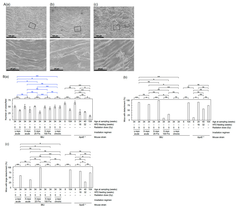
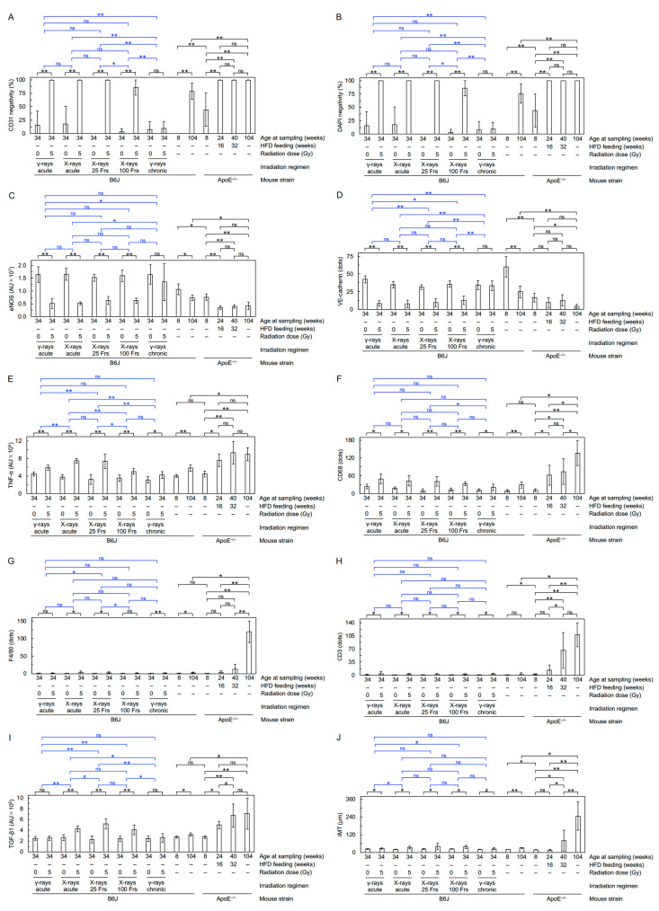
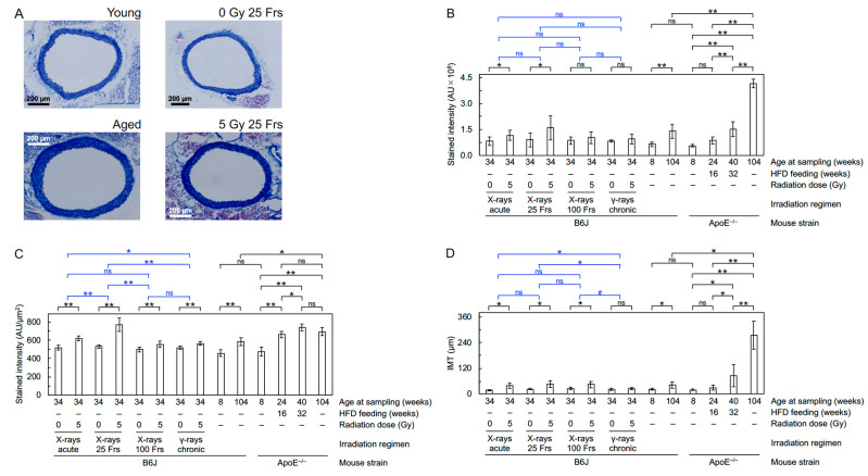
During medical (therapeutic or diagnostic) procedures or in other settings, the circulatory system receives ionizing radiation at various dose rates. Here, we analyzed prelesional changes in the circulatory system of wild-type mice at six months after starting acute, intermittent, or continuous irradiation with 5 Gy of photons. Independent of irradiation regimens, irradiation had little impact on left ventricular function, heart weight, and kidney weight. In the aorta, a single acute exposure delivered in 10 minutes led to structural disorganizations and detachment of the aortic endothelium, and intima-media thickening. These morphological changes were accompanied by increases in markers for profibrosis (TGF-β1), fibrosis (collagen fibers), proinflammation (TNF-α), and macrophages (F4/80 and CD68), with concurrent decreases in markers for cell adhesion (CD31 and VE-cadherin) and vascular functionality (eNOS) in the aortic endothelium. Compared with acute exposure, the magnitude of such aortic changes was overall greater when the same dose was delivered in 25 fractions spread over 6 weeks, smaller in 100 fractions over 5 months, and much smaller in chronic exposure over 5 months. These findings suggest that dose protraction alters vascular damage in the aorta, but in a way that is not a simple function of dose rate.

Keywords: ApoE−/−; C57BL6/J; aged mice; aorta; fibrosis; inflammation; intima-media thickening; ionizing radiation; left ventricular function; vascular damage.


Conflict of interest statement

The authors declare no conflict of interest.

Figures

Figure 1
Figure 1
Morphological changes in the aortic endothelium. (A) Representative FE-SEM images of (a) normal endothelium (young mouse), and (b,c) detachment and large detachment, respectively (6 months after starting irradiation with 5 Gy of X-rays in 25 fractions), all in B6J mice. Boxed areas in the upper panels are shown at higher magnification in the lower panels. Scale bars as indicated. (B) Quantitative analysis for (a) the number of crests/field (7–11 mice/group analyzed except for 4 in aged ApoE−/− mice, Welch’s t-test), and (b,c) percentage of mice with detachment and large detachment, respectively (7–11 mice/group analyzed except for 20 in B6J mice at 6 months after irradiation with 0 Gy or 5 Gy of acute γ-rays and 4 in aged ApoE−/− mice, Fisher’s exact test). For an outline of statistical comparisons, see a footnote in Supplementary Materials. Frs, fractions. The data in Figure 1B(a–c) for the two B6J groups receiving 0 Gy or 5 Gy of acute γ-rays were taken from the 2020 Cancers paper [7]. **, p < 0.001. *, 0.001 ≤ p < 0.05. #, 0.05 ≤ p < 0.1 (marginally significant). ns, p ≥ 0.1 (nonsignificant).
Figure 2
Figure 2
Molecular changes in the aorta. Representative merged images in B6J mice (all at 6 months after starting irradiation) for double immunofluorescence of CD31 with (A) eNOS, (B) VE-cadherin, (C) TNF-α, (D) CD68, (E) F4/80, (F) CD3 or, (G) TGF-β1, with cell nuclei counterstained with DAPI. (A,C,EG) X-rays in 25 fractions. (B,D) X-rays in 100 fractions. Upper panels, 0 Gy. Lower panels, 5 Gy. Boxed areas in the left panels (tiled images) are shown at higher magnification in the right panels. Scale bars as indicated.
Figure 3
Figure 3
Molecular changes in the aorta. Quantitative analysis of immunofluorescence for (A) CD31 negativity, (B) DAPI negativity, (C) eNOS, (D) VE-cadherin, (E) TNF-α, (F) CD68, (G) F4/80, (H) CD3, (I) TGF-β1, and (J) IMT (7–11 mice/group analyzed except for 4 in aged ApoE−/− mice, Welch’s t-test, or Wald test). For clarity, graphs replotted for F4/80, CD3, and IMT in B6J mice only are shown in Figure S6A–C). For an outline of statistical comparisons, see a footnote in Supplementary Materials. AU, arbitrary unit. Frs, fractions. The data in Figure 3A–E,G) for the two B6J groups receiving 0 Gy or 5 Gy of acute γ-rays were taken from the 2020 Cancers paper [7]. Representative images in B6J mice are shown in Figure 2. **, p < 0.001. *, 0.001 ≤ p < 0.05. #, 0.05 ≤ p < 0.1 (marginally significant). ns, p ≥ 0.1 (nonsignificant).
Figure 4
Figure 4
Fibrotic changes in the aorta. (A). Representative images for Masson’s trichrome staining in B6J mice (young, aged, at 6 months after starting irradiation with 0 Gy or 5 Gy of X-rays in 25 fractions). Quantitative analysis for (B) total intensity of aniline blue stain in the entire aortic wall, (C) intensity of aniline blue stain per unit aortic wall area, and (D) IMT (8–10 mice/group analyzed except for 4 in aged ApoE−/− mice, Welch’s t-test). For clarity, a graph replotted for IMT in B6J mice only is shown in Figure S9C. For an outline of statistical comparisons, see a footnote in Supplementary Materials. AU, arbitrary unit. Frs, fractions. **, p < 0.001. *, 0.001 ≤ p < 0.05. #, 0.05 ≤ p < 0.1 (marginally significant). ns, p ≥ 0.1 (nonsignificant).

Similar articles

Cited by

References

    1. Little M.P. Radiation and Circulatory Disease. Mutat. Res. 2016;770:299–318. doi: 10.1016/j.mrrev.2016.07.008. - DOI - PMC - PubMed
    1. Little M.P., Azizova T.V., Hamada N. Low- and Moderate-Dose Non-Cancer Effects of Ionizing Radiation in Directly Exposed Individuals, Especially Circulatory and Ocular Diseases: A Review of the Epidemiology. Int. J. Radiat. Biol. 2021;97:782–803. doi: 10.1080/09553002.2021.1876955. - DOI - PMC - PubMed
    1. Tapio S., Little M.P., Kaiser J.C., Impens N., Hamada N., Georgakilas A.G., Simar D., Salomaa S. Ionizing Radiation-Induced Circulatory and Metabolic Diseases. Environ. Int. 2021;146:106235. doi: 10.1016/j.envint.2020.106235. - DOI - PMC - PubMed
    1. ICRP ICRP Statement on Tissue Reactions/Early and Late Effects of Radiation in Normal Tissues and Organs–Threshold Doses for Tissue Reactions in a Radiation Protection Context: ICRP Publication 118. [(accessed on 28 September 2021)];Ann. ICRP. 2012 41:1–322. doi: 10.1016/j.icrp.2012.02.001. Available online: https://journals.sagepub.com/doi/pdf/10.1177/ANIB_41_1-2. - DOI - DOI - PubMed
    1. Hamada N., Fujimichi Y., Iwasaki T., Fujii N., Furuhashi M., Kubo E., Minamino T., Nomura T., Sato H. Emerging Issues in Radiogenic Cataracts and Cardiovascular Disease. J. Radiat. Res. 2014;55:831–846. doi: 10.1093/jrr/rru036. - DOI - PMC - PubMed

LinkOut - more resources