. 2021 Oct 29;13(21):5446.
doi: 10.3390/cancers13215446.

Enhancement of NK Cell Antitumor Effector Functions Using a Bispecific Single Domain Antibody Targeting CD16 and the Epidermal Growth Factor Receptor

Affiliations

Enhancement of NK Cell Antitumor Effector Functions Using a Bispecific Single Domain Antibody Targeting CD16 and the Epidermal Growth Factor Receptor

Elisa C Toffoli et al. Cancers (Basel). .

Abstract

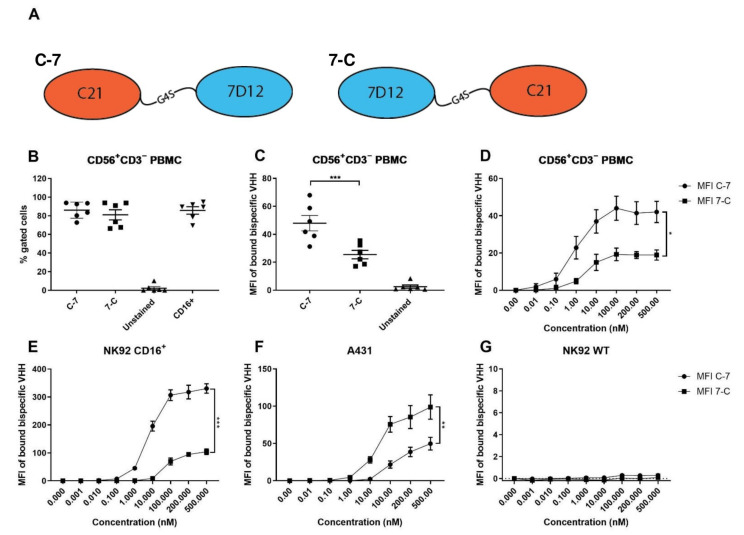
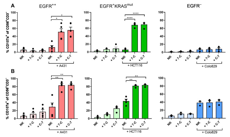
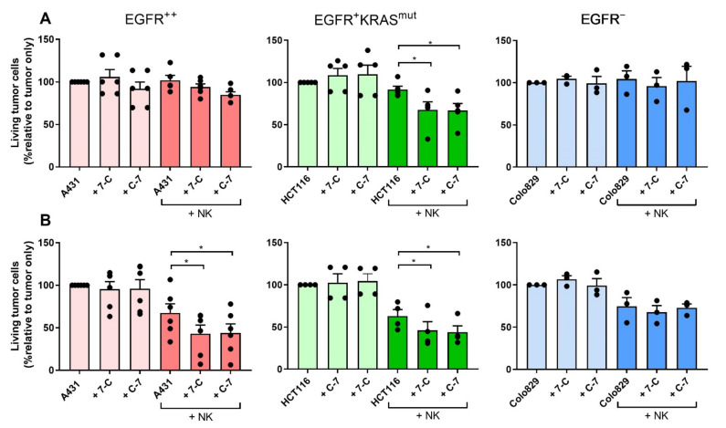
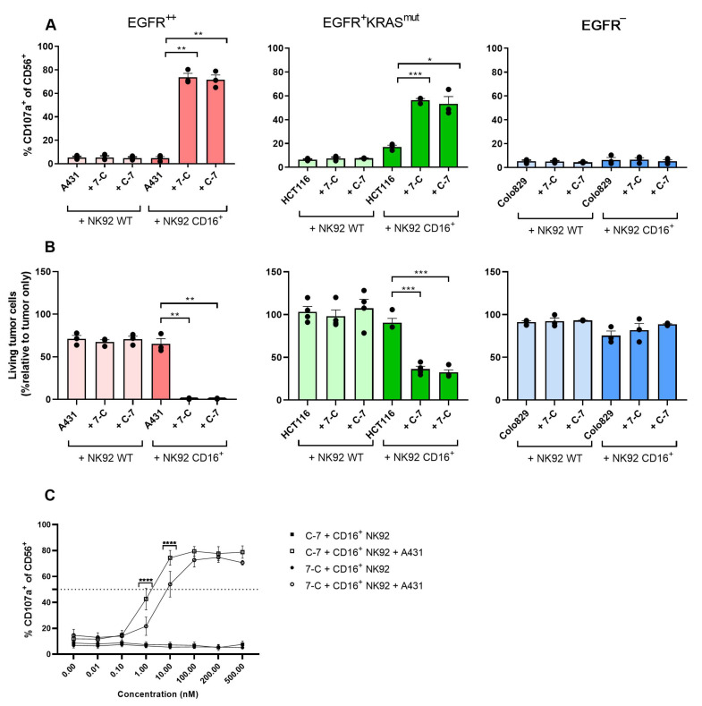
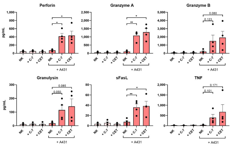
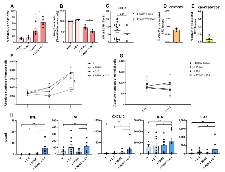
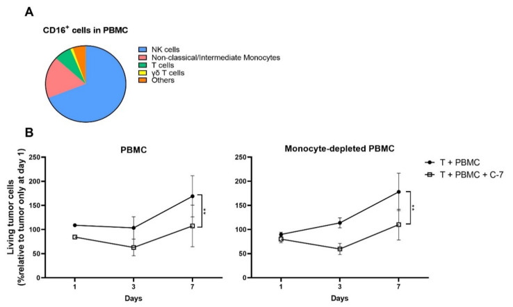
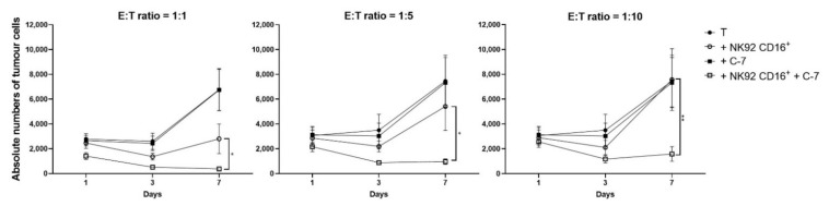
The ability to kill tumor cells while maintaining an acceptable safety profile makes Natural Killer (NK) cells promising assets for cancer therapy. Strategies to enhance the preferential accumulation and activation of NK cells in the tumor microenvironment can be expected to increase the efficacy of NK cell-based therapies. In this study, we show binding of a novel bispecific single domain antibody (VHH) to both CD16 (FcRγIII) on NK cells and the epidermal growth factor receptor (EGFR) on tumor cells of epithelial origin. The bispecific VHH triggered CD16- and EGFR-dependent activation of NK cells and subsequent lysis of tumor cells, regardless of the KRAS mutational status of the tumor. Enhancement of NK cell activation by the bispecific VHH was also observed when NK cells of colorectal cancer (CRC) patients were co-cultured with EGFR expressing tumor cells. Finally, higher levels of cytotoxicity were found against patient-derived metastatic CRC cells in the presence of the bispecific VHH and autologous peripheral blood mononuclear cells or allogeneic CD16 expressing NK cells. The anticancer activity of CD16-EGFR bispecific VHHs reported here merits further exploration to assess its potential therapeutic activity either alone or in combination with adoptive NK cell-based therapeutic approaches.

Keywords: CD16; EGFR; NK cells; bispecific VHH; single domain antibodies.


Conflict of interest statement

Jan Spanholtz is chief scientific officer at Glycostem BV and Hans. J. van der Vliet is chief scientific officer at Lava Therapeutics NV. Tanja de Gruijl is scientific advisor to Lava Therapeutics. Amanda van Vliet is an employee of Glycostem BV. No other conflict of interest is reported.

Figures

Figure 1
Figure 1
Binding characteristics of C-7 and 7-C bispecific VHHs to CD16 and EGFR. (A) Graphical representation of the bispecific VHHs. (B) Percentage of bispecific VHH+ cells (at 100 nM) and CD16+ cells among CD56+CD3 cells in PBMC, n = 6. (C) Median Fluorescence Intensity (MFI) of bound bispecific VHH on CD56+CD3 cells in PBMC at 100 nM, n = 6. (D) MFI of bound bispecific VHH on CD56+CD3 cells in PBMC n = 3. (E) MFI of bound bispecific VHH to CD16+ NK92 n = 3. (F) MFI of bound bispecific VHH to A431 (EGFR++) n = 8; (G) MFI of bound bispecific VHH to CD16EGFR NK92 WT, n = 3. The data are presented as mean ± SEM. Significance is presented as p < 0.05 *, <0.01 **, <0.001 ***. p-values were determined by two-tailed paired t-test (B,C) or two-way ANOVA (D,G). Abbreviations: PBMC = peripheral blood mononuclear cells, C-7 = C21-7D12 bispecific VHH, 7-C = 7D12-C21 bispecific VHH.
Figure 2
Figure 2
CD16-EGFR bispecific VHHs induce degranulation of NK cells in the presence of EGFR-expressing tumor cells. Degranulation of healthy donor-derived NK cells after a 4-h co-culture ±100 nM bispecific VHHs ± tumor cell lines expressing different levels of EGFR: A431 (EGFR++), HCT116 (EGFR+RASmut), Colo829 (EGFR). (A) Resting NK cells were used as effectors; (B) NK cells were pre-activated overnight with IL-2 and IL-15. Degranulation was determined by assessing the percentage of CD107a+ cells. E:T ratio: 1:1. The bars represent mean ± SEM. Significance is presented as p < 0.05 *, <0.01 **, <0.0001 ****. A431 n = 5 (A,B); HCT116 n = 4 (A,B); Colo829 n = 3 (A,B). p-values were determined by two-tailed paired t-test. Abbreviations: C-7 = C21-7D12 bispecific VHH, 7-C = 7D12-C21 bispecific VHH.
Figure 3
Figure 3
CD16-EGFR bispecific VHHs induce lysis of EGFR expressing tumor cells in the presence of NK cells. Cytotoxicity exerted by healthy donor-derived NK cells after a 24-h co-culture ±100 nM bispecific VHHs ± tumor cell lines expressing different levels of EGFR: A431 (EGFR++), HCT116 (EGFR+RASmut), Colo829 (EGFR). (A) Resting NK cells were used as effectors; (B) NK cells were pre-activated overnight with IL-2 and IL-15. Cytotoxicity was determined by assessing the relative percentage of living tumor cells compared to the tumor-alone condition. E:T ratio: 1:1. The bars represent mean ± SEM. Significance is presented as p < 0.05 *. A431 n = 6 (A,B); HCT116 n = 5 (A) and n = 4 (B); Colo829 n = 3 (A,B). p-values were determined by a two-tailed paired t-test. Abbreviations: C-7 = C21-7D12 bispecific VHH, 7-C = 7D12-C21 bispecific VHH.
Figure 4
Figure 4
CD16-EGFR bispecific VHHs induce degranulation of CD16+ NK92 cells and cytotoxicity against EGFR-expressing tumor cells. Degranulation (A) and cytotoxicity (B) of NK92 WT and NK92 CD16+ co-cultured ±100 nM bispecific VHHs ± tumor cell lines expressing different levels of EGFR: A431 (EGFR++), HCT116 (EGFR+RASmut), Colo829 (EGFR). Degranulation was determined after 4 h by assessing the percentage of CD107a+ cells. Cytotoxicity was determined after 24 h by assessing the relative percentage of living tumor cells compared to the tumor-alone condition. E:T ratio: 1:1. The bars represent mean ± SEM. A431 n = 3 (A,B); HCT116 n = 3 (A) and n = 4 (B); Colo829 n = 3 (A,B). (C) Degranulation of NK92 CD16+ after 4-h co-culture with A431 ± concentration range of the bispecific VHHs. Degranulation was assessed by determining the percentage of CD107a+ cells. E:T ratio: 1:1. The data are presented as mean ± SEM. n = 4. Significance is presented as p < 0.05 *, <0.01 **, <0.001 ***, <0.0001 ****. p-values were determined by two-tailed paired t-test (A,B) or with two-way ANOVA with Bonferroni multiple comparison analysis (C). Abbreviations: C-7 = C21-7D12 bispecific VHH, 7-C = 7D12-C21 bispecific VHH.
Figure 5
Figure 5
The C-7 bispecific VHH and cetuximab trigger NK cell secretion of pro-inflammatory and cytotoxic mediators. Cytometry Bead Array performed on supernatant of overnight-activated healthy donor-derived NK cells ± A431 ± 100 nM bispecific VHHs ±5 μg/mL cetuximab. E:T ratio 1:1. n = 4. The data are presented as mean ± SEM. Significance is indicated or presented as p < 0.05 *, <0.01 **. p-values are determined by two-tailed paired t-test. Abbreviations: C-7 = C21-7D12 bispecific VHH, CET = cetuximab.
Figure 6
Figure 6
Activity of the bispecific C-7 VHH using metastatic CRC patient-derived PBMC and dissociated tumor samples. (A) Degranulation of NK cells (defined as CD45+CD56+CD3 cells) and (B) cytotoxicity of A431 tumor cells after a 4-h (degranulation) and 24-h (cytotoxicity) co-culture of monocyte-depleted PBMC derived from patients with stage II/III CRC pre-activated overnight with IL-2 and IL-15 ± A431 (EGFR++) ±100 nM C-7. E:T ratio 4:1. Degranulation was determined by assessing the percentage of NK cells expressing CD107a+. Cytotoxicity was determined by assessing the relative percentage of living tumor cells compared to the tumor-alone control. n = 5 (A), n = 4 (B). (C) EGFR MFI of Epcam+CD45 tumor cells in dissociated CRC peritoneal metastatic lesions (n = 15) and EpcamdimCD45 epithelial cells, in (non-malignant) peritoneal tissue (n = 3). CD56+CD3 (D) and CD16+CD56+CD3 (E) percentage of CD45+ cells in dissociated CRC peritoneal metastatic lesions, n = 6. (F) Absolute number of Epcam+CD45 tumor cells from dissociated CRC peritoneal metastatic lesions cultured ± autologous PBMC (E:T ratio 5:1) ±100 nM C-7 for 1, 3, and 7 days. n = 8. The significance level refers to the conditions: “T+PBMC” versus “T+PBMC+C-7”. (G) Absolute number of EpcamdimCD45 cells from dissociated normal (non-malignant) peritoneal tissue of patients with metastatic CRC ± autologous PBMC (E:T ratio 5:1) ±100 nM C-7, incubated for 1 and 3 days. n = 3. (H) Cytometry Bead Array performed on supernatant of dissociated CRC peritoneal metastases cultured for 24 h ± autologous PBMC (E:T ratio 5:1) ± 100 nM C-7. n = 9. The data are presented as mean ± SEM. Significance is presented as p < 0.05 *, <0.01 **, <0.001 ***. No p-values are mentioned in case p > 0.05. p-values are determined by two-tailed paired t-test (A,B); Mann–Whitney test (C); two-way ANOVA with Dunnett’s multiple comparison test (F,G); one-way ANOVA with Dunnett’s multiple comparison test (H: IL-6); Friedman test with Dunn’s multiple comparison test (H: IFNγ, TNF, CXCL10, IL-10). Abbreviations: T = tumor, C-7 = C21-7D12 bispecific VHH, PBMC = peripheral blood mononuclear cells.
Figure 7
Figure 7
Tumor growth control mediated by the bispecific C-7 VHH in co-cultures of metastatic CRC cells and autologous PBMC is independent of effects on non-classical/intermediate monocytes. (A) Distribution of CD16+ cells in PBMC from patients with peritoneal CRC metastasis. n = 7. (B) Dissociated CRC peritoneal metastatic lesions ±100 nM C-7 ± autologous PBMC or monocyte depleted autologous PBMC. E:T ratio: 5:1. n = 4. Incubated for 1, 3, and 7 days. Data are presented as mean ± SEM. Significance is presented as p < 0.01 **. p-values were determined by two-way ANOVA with Tukey’s multiple comparison test (B). Abbreviations: T = tumor, C-7 = C21-7D12 bispecific VHH.
Figure 8
Figure 8
The bispecific C-7 VHH triggers CD16+ NK92 cells to control tumor growth using patient metastatic CRC cells. Dissociated CRC peritoneal metastatic samples ± NK92 CD16+ ±100 nM C-7 were cultured for 1, 3, and 7 days at multiple E:T ratios, i.e., 1:1 n = 6, 1:5 n = 5, 1:10 n = 5. The significance levels refer to the conditions: “T+NK92 CD16+” versus “T+NK92 CD16++C-7”. Data are presented as mean ± SEM. Significance is presented as p < 0.05 *, <0.01 **. p-values were determined by two-way ANOVA with Dunnett’s multiple comparison analysis. Abbreviations: T = tumor, C-7 = C21-7D12 bispecific VHH.

Similar articles

Cited by

References

    1. Vivier E., Tomasello E., Baratin M., Walzer T., Ugolini S. Functions of Natural Killer Cells. Nat. Immunol. 2008;9:503–510. doi: 10.1038/ni1582. - DOI - PubMed
    1. Miller J.S. The Biology of Natural Killer Cells in Cancer, Infection, and Pregnancy. Exp. Hematol. 2001;29:1157–1168. doi: 10.1016/S0301-472X(01)00696-8. - DOI - PubMed
    1. Cooper M.A., Fehniger T.A., Caligiuri M.A. The Biology of Human Natural Killer-Cell Subsets. Trends Immunol. 2001;22:633–640. doi: 10.1016/S1471-4906(01)02060-9. - DOI - PubMed
    1. Campbell K.S., Hasegawa J. Natural Killer Cell Biology: An Update and Future Directions. J. Allergy Clin. Immunol. 2013;132:536–544. doi: 10.1016/j.jaci.2013.07.006. - DOI - PMC - PubMed
    1. Terunuma H., Deng X., Dewan Z., Fujimoto S., Yamamoto N. Potential Role of NK Cells in the Induction of Immune Responses: Implications for NK Cell-Based Immunotherapy for Cancers and Viral Infections. Int. Rev. Immunol. 2008;27:93–110. doi: 10.1080/08830180801911743. - DOI - PubMed