Review
. 2021 Oct 29;13(21):5455.
doi: 10.3390/cancers13215455.

The Modes of Dysregulation of the Proto-Oncogene T-Cell Leukemia/Lymphoma 1A

Affiliations
Review

The Modes of Dysregulation of the Proto-Oncogene T-Cell Leukemia/Lymphoma 1A

Johanna Stachelscheid et al. Cancers (Basel). .

Abstract

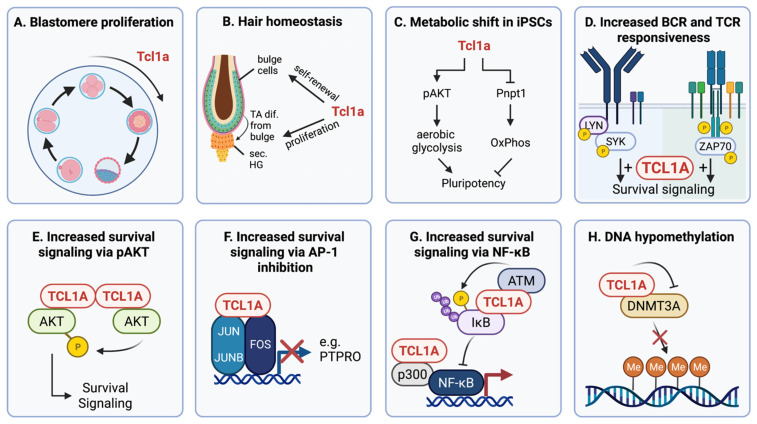
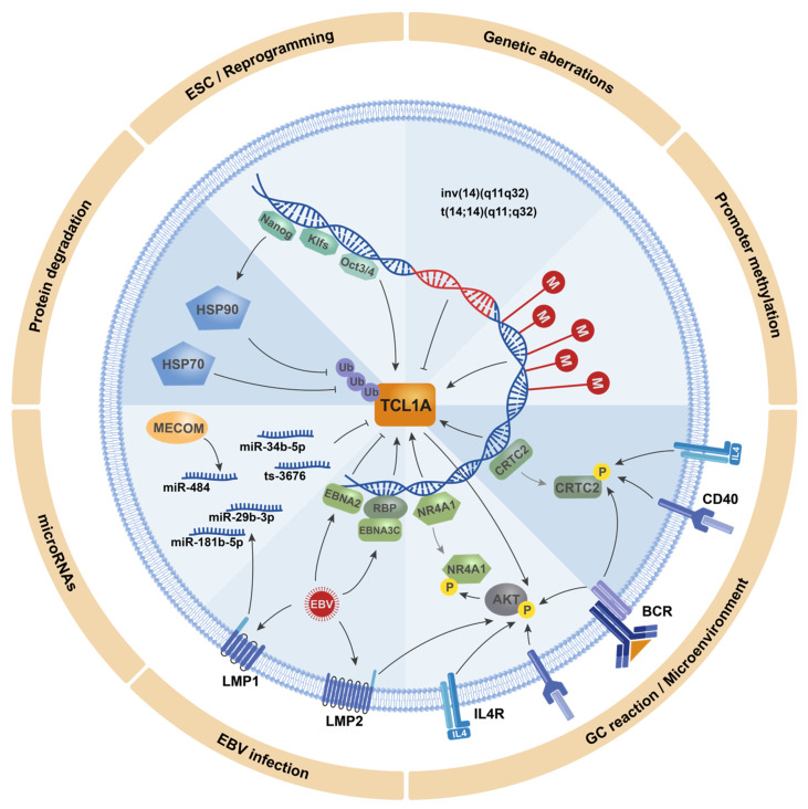
Incomplete biological concepts in lymphoid neoplasms still dictate to a large extent the limited availability of efficient targeted treatments, which entertains the mostly unsatisfactory clinical outcomes. Aberrant expression of the embryonal and lymphatic TCL1 family of oncogenes, i.e., the paradigmatic TCL1A, but also TML1 or MTCP1, is causally implicated in T- and B-lymphocyte transformation. TCL1A also carries prognostic information in these particular T-cell and B-cell tumors. More recently, the TCL1A oncogene has been observed also in epithelial tumors as part of oncofetal stemness signatures. Although the concepts on the modes of TCL1A dysregulation in lymphatic neoplasms and solid tumors are still incomplete, there are recent advances in defining the mechanisms of its (de)regulation. This review presents a comprehensive overview of TCL1A expression in tumors and the current understanding of its (dys)regulation via genomic aberrations, epigenetic modifications, or deregulation of TCL1A-targeting micro RNAs. We also summarize triggers that act through such transcriptional and translational regulation, i.e., altered signals by the tumor microenvironment. A refined mechanistic understanding of these modes of dysregulations together with improved concepts of TCL1A-associated malignant transformation can benefit future approaches to specifically interfere in TCL1A-initiated or -driven tumorigenesis.

Keywords: BPDCN; CLL; T-PLL; TCL1 oncogenes; kinase signaling; lymphoma.


Conflict of interest statement

The authors declare no conflict of interest.

Figures

Figure 1
Figure 1
TCL1A functions as a pleiotropic adapter molecule in stemness and survival signaling. Note that “roles” and “modes of actions” at various levels (e.g., cell function, impacted pathway, concise molecular interaction) are highlighted and are in part artificially separated (i.e., E part of C and D): (A) in murine blastomeres, Tcl1a is important in early proliferation, as Tcl1a−/− mice show a block of blastomere development at the 8-cell stage [9]; (B) Tcl1a regulated hair growth, as shown by hair loss in Tcl1a−/− mice. Tcl1a is expressed in the bulge cells (stem cell niche) and in the secondary hair germ/transit-amplifying (TA) cells (proliferative structure) during the catagen–telogen (resting phase) transition and early anagen stage (regeneration phase). In Tcl1a−/− mice, the bulge cells show reduced expression of the stem cell marker CD34. Furthermore, a Tcl1a knockout led to reduced proliferation of TA cells, needed for new hair formation [59]; (C) upregulation of Tcl1a leads to metabolic shifts toward aerobic glycolysis via activation of Akt and repression of Pnpt1, thereby contributing to pluripotency of induced pluripotent stem cells (iPSCs) [60]; (D) in cells of chronic lymphocytic leukemia (CLL) and T-cell prolymphocytic leukemia (T-PLL), TCL1A increases the responsiveness to B-cell receptor (BCR) and T-cell receptor (TCR) stimulation, respectively, by a kinase activating effect [24,26,61]; (E) interaction of the TCL1A homodimer with AKT molecules leads to augmented trans-phosphorylation and catalytic activity of the oncogenic Ser/Thr kinase AKT, resulting in increased survival signaling [62]; (F) the interaction of TCL1A with AP-1 components—namely, JUN, JUNB, and FOS, leads to impaired AP-1 signaling and thereby sustained anti-apoptotic signals [11]; (G) TCL1A interacts with IκB and mediates its phosphorylation via ATM, leading to its subsequent ubiquitination-dependent degradation. Inhibition of this negative regulator IκB causes increased NF-κB signaling, which is additionally strengthened by the TCL1A-p300 interaction [11]; (H) physical interaction of TCL1A with DNMT3A reduces the methyltransferase activity of DNMT3A, which leads to a higher number of hypomethylated genomic regions [63], which is implicated in the pathogenesis of CLL [64]. This figure was created using BioRender.com (accessed on 22 October 2021).
Figure 2
Figure 2
Schematic overview of the different modes of TCL1A regulation (clockwise categories). ESC/Reprogramming: In murine embryonic stem cells (ESCs) and induced pluripotent stem cells (iPSCs), Tcl1a expression is mediated by the transcription factors Nanog [75,76,81], Klf2/4/5 [60,82], and Oct3/4 [70]. Genetic aberrations: In T-cell prolymphocytic leukemia (T-PLL), an inversion or translocation of the TCL1A gene on chromosome 14 positions its locus under control by highly active regulatory regions of T-cell receptor genes. This prevents TCL1A’s post-thymic silencing and causes its constitutive expression [1]. Promoter methylation: During T-cell and B-cell development and maturation, a protracted epigenetic silencing of TCL1A expression is likely. B-cell lines have shown three different patterns of promoter methylation, that might reflect a successive increase in methylation along B-cell differentiation [83]. GC reaction/microenvironment: Signals via the BCR and/or via IL4R and CD40 ligation lead to phosphorylation and nuclear exclusion of CRTC2 [84] and NR4A1 [85], thereby repressing transcriptional activation of TCL1A. EBV infections: These signals by the microenvironment can also be mimicked by the Epstein–Barr virus (EBV) proteins LMP1 and LMP2, leading to repression of TCL1A [86,87]. Furthermore, the EBV protein EBNA2 represses [87], whereas EBNA3C increases, TCL1A expression [88]. MiRNAs: At the post-transcriptional level, TCL1A is regulated by several microRNAs (miRs), whose expressions are deregulated via co-deletion at 13q [89] and 17p [90], the EBV protein LMP1 [86], and the protein MECOM [29]. Protein degradation: Integrity of the TCL1A protein is regulated via the chaperones HSP70 [91] and HSP90 [76] that protect TCL1A from ubiquitination and subsequent degradation. Expression of HSP90 is also regulated by the stem cell factor Nanog, which thereby mediates a bimodal regulation of TCL1A at the gene and protein level [76]. Grey arrows: dissociation from the TCL1A promoter.

References

    1. Virgilio L., Narducci M.G., Isobe M., Billips L.G., Cooper M.D., Croce C.M., Russo G. Identification of the TCL1 gene involved in T-cell malignancies. Proc. Natl. Acad. Sci. USA. 1994;91:12530–12534. doi: 10.1073/pnas.91.26.12530. - DOI - PMC - PubMed
    1. Russo G., Isobe M., Gatti R., Finan J., Batuman O., Huebner K., Nowell P.C., Croce C.M. Molecular analysis of a t(14;14) translocation in leukemic T-cells of an ataxia telangiectasia patient. Proc. Natl. Acad. Sci. USA. 1989;86:602–606. doi: 10.1073/pnas.86.2.602. - DOI - PMC - PubMed
    1. Virgilio L., Isobe M., Narducci M.G., Carotenuto P., Camerini B., Kurosawa N., Abbas-ar-Rushdi, Croce C.M., Russo G. Chromosome walking on the TCL1 locus involved in T-cell neoplasia. Proc. Natl. Acad. Sci. USA. 1993;90:9275–9279. doi: 10.1073/pnas.90.20.9275. - DOI - PMC - PubMed
    1. Teitell M.A. The TCL1 family of oncoproteins: Co-activators of transformation. Nat. Rev. Cancer. 2005;5:640–648. doi: 10.1038/nrc1672. - DOI - PubMed
    1. Pekarsky Y., Hallas C., Isobe M., Russo G., Croce C.M. Abnormalities at 14q32.1 in T cell malignancies involve two oncogenes. Proc. Natl. Acad. Sci. USA. 1999;96:2949–2951. doi: 10.1073/pnas.96.6.2949. - DOI - PMC - PubMed

LinkOut - more resources