

Performance of Contrast-Enhanced Ultrasound in Thyroid Nodules: Review of Current State and Future Perspectives
- PMID: 34771632
- PMCID: PMC8582579
- DOI: 10.3390/cancers13215469
Performance of Contrast-Enhanced Ultrasound in Thyroid Nodules: Review of Current State and Future Perspectives
Abstract
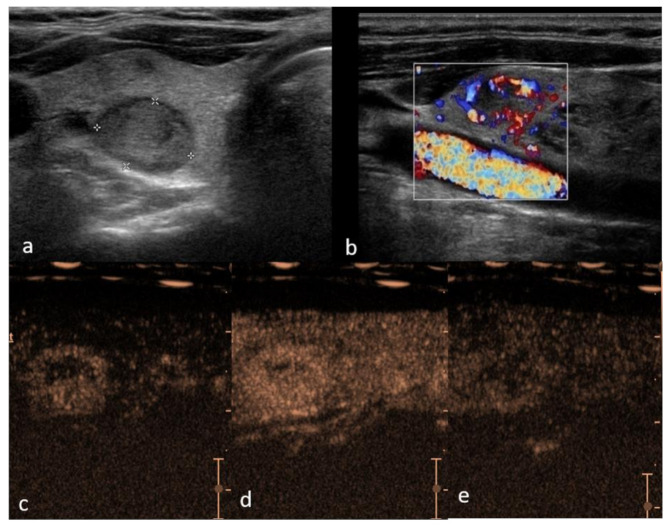
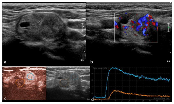
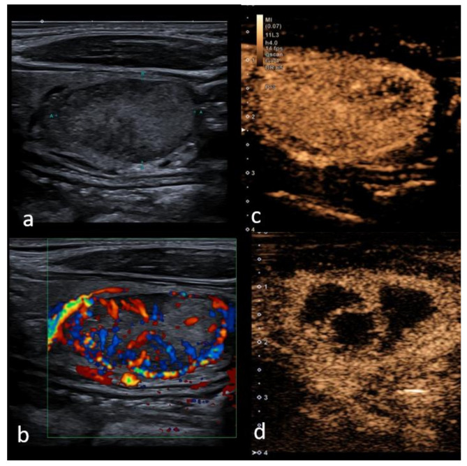
Ultrasound has been established as a baseline imaging technique for thyroid nodules. The main advantage of adding CEUS is the ability to assess the sequence and intensity of vascular perfusion and hemodynamics in the thyroid nodule, thus providing real-time characterization of nodule features, considered a valuable new approach in the determination of benign vs. malignant nodules. Original studies, reviews and six meta-analyses were included in this article. A total of 624 studies were retrieved, and 107 were included in the study. As recognized for thyroid nodule malignancy risk stratification by US, for acceptable accuracy in malignancy a combination of several CEUS parameters should be applied: hypo-enhancement, heterogeneous, peripheral irregular enhancement in combination with internal enhancement patterns, and slow wash-in and wash-out curve lower than in normal thyroid tissue. In contrast, homogeneous, intense enhancement with smooth rim enhancement and "fast-in and slow-out" are indicative of the benignity of the thyroid nodule. Even though overlapping features require standardization, with further research, CEUS may achieve reliable performance in detecting or excluding thyroid cancer. It can also play an operative role in guiding ablation procedures of benign and malignant thyroid nodules and metastatic lymph nodes, and providing accurate follow-up imaging to assess treatment efficacy.
Keywords: contrast-enhanced ultrasound (CEUS); follicular thyroid cancer; papillary thyroid cancer; thyroid cancer; thyroid nodules.
Conflict of interest statement
Maija Radzina has received the speaker honoraria from Bracco and Canon, other co-authors have nothing to disclose.
Figures
References
-
- Haugen B.R., Alexander E.K., Bible K.C., Doherty G.M., Mandel S.J., Nikiforov Y.E., Pacini F., Randolph G.W., Sawka A.M., Schlumberger M., et al. 2015 American Thyroid Association Management Guidelines for Adult Patients with Thyroid Nodules and Differentiated Thyroid Cancer: The American Thyroid Association Guidelines Task Force on Thyroid Nodules and Differentiated Thyroid Cancer. Thyroid. 2016;26:1–133. doi: 10.1089/thy.2015.0020. - DOI - PMC - PubMed
-
- Pang T., Huang L., Deng Y., Wang T., Chen S., Gong X., Liu W. Logistic Regression Analysis of Conventional Ultrasonography, Strain Elastosonography, and Contrast-Enhanced Ultrasound Characteristics for the Differentiation of Benign and Malignant Thyroid Nodules. PLoS ONE. 2017;12:e0188987. doi: 10.1371/journal.pone.0188987. - DOI - PMC - PubMed
Publication types
LinkOut - more resources
Full Text Sources
Medical
Research Materials
