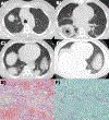
Management of Pulmonary Mucormycosis After Orthotopic Heart Transplant: A Case Series
- PMID: 34772489
- PMCID: PMC9034368
- DOI: 10.1016/j.transproceed.2021.09.034
Management of Pulmonary Mucormycosis After Orthotopic Heart Transplant: A Case Series
Abstract
Invasive pulmonary mucormycosis is a potentially fatal infection that can occur in immunosuppressed patients such as those who have undergone orthotopic heart transplant (OHT). High-dose intravenous antifungal agents, including amphotericin B, are generally accepted as the first-line medical treatment, with prompt surgical resection of lesions if feasible. The body of evidence guiding treatment decisions, however, is sparse, particularly regarding adjustment of immunosuppression during acute infection and long-term recovery. We present 2 cases of patients with pulmonary mucormycosis occurring within the first 6 months after OHT, both of whom successfully recovered after appropriate medical and surgical treatment, and we highlight differences in immunosuppression management strategies for this life-threatening condition.
Copyright © 2021 Elsevier Inc. All rights reserved.
Figures


References
-
- Jeong W, Keighley C, Wolfe R, et al. The epidemiology and clinical manifestations of mucormycosis: a systematic review and meta-analysis of case reports. Clin Microbiol Infect 2019;25:26–34. - PubMed
-
- Bardwell J, Youseffi B, Marquez J, Zangeneh TT, Al-Obaidi M. Pulmonary mucormycosis in a heart transplant patient. Am J Med 2020;133:e524–5. - PubMed
-
- Nokes BT, Pajaro O, Stephen J, et al. Monster lung cavity in a heart transplant recipient. Heart Surg Forum 2018;21:E072–4. - PubMed
-
- Margoles L, DeNofrio D, Patel AR, et al. Disseminated mucormycosis masquerading as rejection early after orthotopic heart transplantation. Transpl Infect Dis 2018;20:e12820. - PubMed
MeSH terms
Substances
Grants and funding
LinkOut - more resources
Full Text Sources
Medical

