Lung ultrasound volume sweep imaging for respiratory illness: a new horizon in expanding imaging access
- PMID: 34772730
- PMCID: PMC8593737
- DOI: 10.1136/bmjresp-2021-000919
Lung ultrasound volume sweep imaging for respiratory illness: a new horizon in expanding imaging access
Abstract
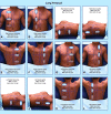
Background: Respiratory illness is a leading cause of morbidity in adults and the number one cause of mortality in children, yet billions of people lack access to medical imaging to assist in its diagnosis. Although ultrasound is highly sensitive and specific for respiratory illness such as pneumonia, its deployment is limited by a lack of sonographers. As a solution, we tested a standardised lung ultrasound volume sweep imaging (VSI) protocol based solely on external body landmarks performed by individuals without prior ultrasound experience after brief training. Each step in the VSI protocol is saved as a video clip for later interpretation by a specialist.
Methods: Dyspneic hospitalised patients were scanned by ultrasound naive operators after 2 hours of training using the lung ultrasound VSI protocol. Separate blinded readers interpreted both lung ultrasound VSI examinations and standard of care chest radiographs to ascertain the diagnostic value of lung VSI considering chest X-ray as the reference standard. Comparison to clinical diagnosis as documented in the medical record and CT (when available) were also performed. Readers offered a final interpretation of normal, abnormal, or indeterminate/borderline for each VSI examination, chest X-ray, and CT.
Results: Operators scanned 102 subjects (0-89 years old) for analysis. Lung VSI showed a sensitivity of 93% and a specificity of 91% for an abnormal chest X-ray and a sensitivity of 100% and a specificity of 93% for a clinical diagnosis of pneumonia. When any cases with an indeterminate rating on chest X-ray or ultrasound were excluded (n=38), VSI lung ultrasound showed 92% agreement with chest X-ray (Cohen's κ 0.83 (0.68 to 0.97, p<0.0001)). Among cases with CT (n=21), when any ultrasound with an indeterminate rating was excluded (n=3), there was 100% agreement with VSI.
Conclusion: Lung VSI performed by previously inexperienced ultrasound operators after brief training showed excellent agreement with chest X-ray and high sensitivity and specificity for a clinical diagnosis of pneumonia. Blinded readers were able to identify other respiratory diseases including pulmonary oedema and pleural effusion. Deployment of lung VSI could benefit the health of the global community.
Keywords: pleural effusion; pneumonia; pulmonary edema; teleultrasound; ultrasound.
© Author(s) (or their employer(s)) 2021. Re-use permitted under CC BY-NC. No commercial re-use. See rights and permissions. Published by BMJ.
Conflict of interest statement
Competing interests: Benjamin Castaneda has financial stake in the company Medical Innovation and Technology which seeks to bring ultrasound to rural communities. The other authors declare no conflict of interest.
Figures


References
-
- Rudan I, Tomaskovic L, Boschi-Pinto C, et al. . Global estimate of the incidence of clinical pneumonia among children under five years of age. Bull World Health Organ 2004;82:895–903. doi:/S0042-96862004001200005 - PMC - PubMed
-
- Forum of International Respiratory Societies . The global impact of respiratory disease -. Second Edition, 2017.
-
- Avendaño Carvajal L, Perret Pérez C. Epidemiology of Respiratory Infections. In: Pediatric respiratory diseases: a comprehensive textbook. Cham: Springer International Publishing, 2020: 263–72.
Publication types
MeSH terms
LinkOut - more resources
Full Text Sources
Medical
Research Materials
