. 2021 Nov 16;118(46):e2102206118.
doi: 10.1073/pnas.2102206118.

Csi-let-7a-5p delivered by extracellular vesicles from a liver fluke activates M1-like macrophages and exacerbates biliary injuries

Affiliations

Csi-let-7a-5p delivered by extracellular vesicles from a liver fluke activates M1-like macrophages and exacerbates biliary injuries

Chao Yan et al. Proc Natl Acad Sci U S A. .

Abstract

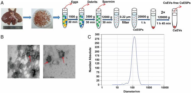
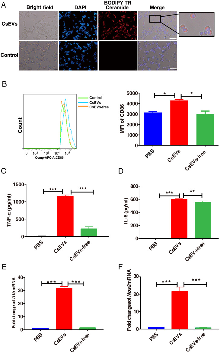
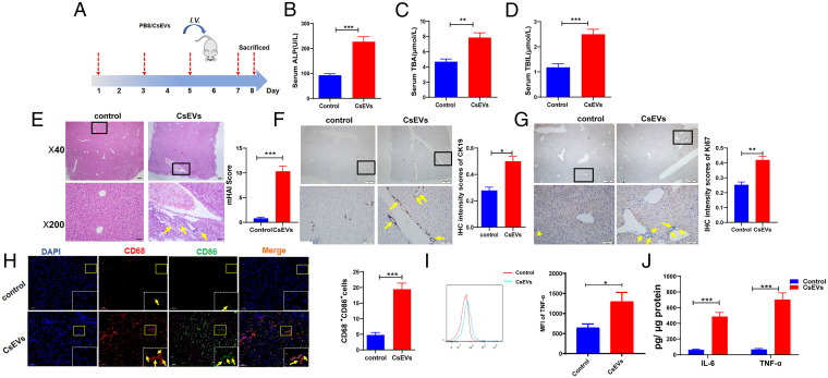
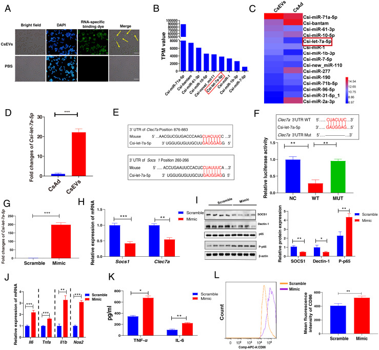
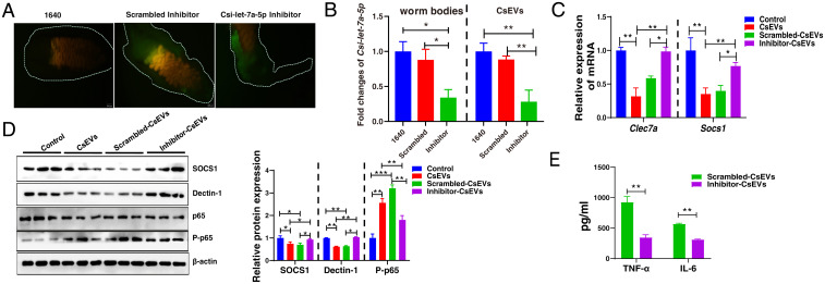
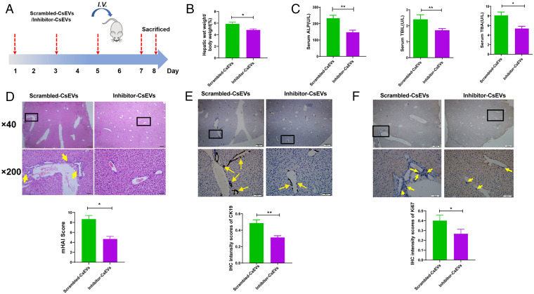
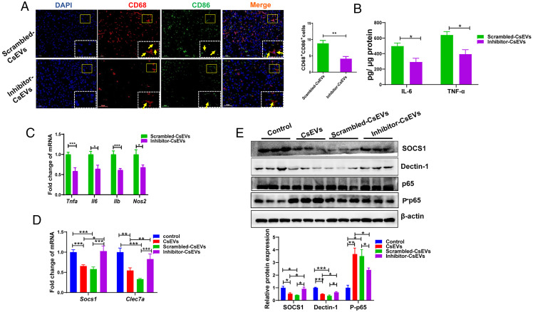
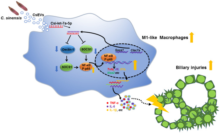
Chronic infection with liver flukes (such as Clonorchis sinensis) can induce severe biliary injuries, which can cause cholangitis, biliary fibrosis, and even cholangiocarcinoma. The release of extracellular vesicles by C. sinensis (CsEVs) is of importance in the long-distance communication between the hosts and worms. However, the biological effects of EVs from liver fluke on biliary injuries and the underlying molecular mechanisms remain poorly characterized. In the present study, we found that CsEVs induced M1-like activation. In addition, the mice that were administrated with CsEVs showed severe biliary injuries associated with remarkable activation of M1-like macrophages. We further characterized the signatures of miRNAs packaged in CsEVs and identified a miRNA Csi-let-7a-5p, which was highly enriched. Further study showed that Csi-let-7a-5p facilitated the activation of M1-like macrophages by targeting Socs1 and Clec7a; however, CsEVs with silencing Csi-let-7a-5p showed a decrease in proinflammatory responses and biliary injuries, which involved in the Socs1- and Clec7a-regulated NF-κB signaling pathway. Our study demonstrates that Csi-let-7a-5p delivered by CsEVs plays a critical role in the activation of M1-like macrophages and contributes to the biliary injuries by targeting the Socs1- and Clec7a-mediated NF-κB signaling pathway, which indicates a mechanism contributing to biliary injuries caused by fluke infection. However, molecules other than Csi-let-7a-5p from CsEVs that may also promote M1-like polarization and exacerbate biliary injuries are not excluded.

Keywords: Clonorchis sinensis; Csi-let-7a-5p; M1 macrophage; biliary injuries; extracellular vesicles.


Conflict of interest statement

The authors declare no competing interest.

Figures

Fig. 1.
Fig. 1.
Identification and characterization of EVs secreted by adults of C. sinensis. (A) Strategy of isolating CsEVs using differential centrifugation. (B) Morphology and structure of CsEVs released in culture medium by adult C. sinensis for 24 h were visualized by TEM (red arrow). (C) The size and distribution of CsEVs were determined using a nanoparticle tracking analysis (NTA).
Fig. 2.
Fig. 2.
CsEVs induce the activation of M1-like macrophages. (A) The red dye-labeled CsEVs were internalized by RAW 264.7 after treatment for 12 h. (Scale bars, 50 μm.) (B) The expression of CD86 in F4/80+CD11b+ BMDMs after treatment with PBS, CsEVs (32 μg/mL), or CsEVs-free (32 μg/mL) for 24 h. (C and D) The secretions of TNF-α (C) and IL-6 (D) produced by the BMDMs after treatment with PBS, CsEVs (32 μg/mL), or CsEVs-free (32 μg/mL) for 24 h as determined by ELISA. (E and F) The relative expression of Il1b (encoding IL-1β) and Nos2 (encoding iNOS) in the BMDMs after treatment with PBS, CsEVs (32 μg/mL) or CsEVs-free (32 μg/mL) for 24 h as determined by qPCR. The values were expressed as mean ± SEM. Compared with indicated groups, *P < 0.05, **P < 0.01, ***P < 0.001.
Fig. 3.
Fig. 3.
CsEVs cause severe biliary injuries. (A) The study design. The mice that were intravenously injected with CsEVs (0.5 mg/mL, ∼100 μL) or PBS (100 μL) at the indicated time. (BD) The levels of serum indicators for biliary injuries, such as ALP (B), TBA (C), and TBIL (D) in the mice treated by CsEVs or PBS. (E) H&E staining showing histological changes (Left) was evaluated using mHAI scoring (Right) in the liver of mice treated with CsEVs or PBS; yellow arrows showed infiltered immune cells. (F) The expression of CK19 (yellow arrows) in the liver of mice treated with CsEVs or PBS by IHC staining. (G) The expression of Ki67 (yellow arrows) in the liver of mice treated with CsEVs or PBS by IHC staining. (H) The proportion of CD86+CD68+macrophages (M1-like, yellow arrows) in the livers of mice that were hydrodynamically injected with CsEVs or PBS as determined by immunofluorescence assay. (I) TNF-α–producing macrophages (CD11b+F4/80+) in livers of these mice were detected using flow cytometry. (J) The levels of IL-6 and TNF-α in the livers of mice were determined by ELISA. The values were expressed as mean ± SEM. Compared with indicated groups, *P < 0.05, **P < 0.01, ***P < 0.001.
Fig. 4.
Fig. 4.
Characterization of enriched miRNAs profiles in CsEVs and identification of the targets of Csi-let-7a-5p packaged by CsEVs. (A) CsEVs delivering miRNAs labeled with the green dyes were found within the macrophages as shown by the fluorescence microscope (scale bars, 50 μm). The yellow arrow shows the internalized miRNA in the macrophage cell line RAW 264.7. (B) The top nine most abundant miRNAs (>1,000 TPM values) in CsEVs were identified using high-throughput sequencing. (C) The comparison of the abundances of miRNAs (>100 TPM) between CsEVs and adult worms showed by the heatmap. (D) The relative expression of Csi-let-7a-5p in the CsEVs and adult worms were determined by qPCR. (E) The potential binding sites of Csi-let-7a-5p in 3′UTR of Clec7a and Socs1 were predicted using the intersection of TargetScan, MiRanda, and PicTar databases. (F) The luciferase activities were measured in the 293T cells that were transfected with either a pmiReport empty vector control (NC), Clec7a-3′UTR (full length, WT), or deletion of Csi-let-7a-5p binding site on Clec7a 3′UTR (MUT) report gene plasmid for 24 h. (G) The levels of Csi-let-7a-5p in macrophages after Csi-let-7a-5p mimic were transfected for 24 h using qPCR. (H) The relative mRNA expression of Socs1 and Clec7a in the macrophages that were transfected with Csi-let-7a-5p mimic or scramble for 24 h. (I) The levels of SOCS1, Dectin-1 (encoded by Clec7a), and P-p65 in macrophages that were transfected with Csi-let-7a-5p mimic or scramble for 24 h. (J) The relative mRNA expression of Il6, Tnfa, Il1b, and Nos2 in the macrophage that was transfected with Csi-let-7a-5p mimic or scramble for 24 h. (K) The secretion of TNF-α and IL-6 in the cultures of macrophages that were transfected with Csi-let-7a-5p mimic or scramble for 24 h. (L) The mean fluorescence intensity of CD86 in macrophages that were transfected with Csi-let-7a-5p mimic or scramble for 24 h using flow cytometry. The values were expressed as mean ± SEM. Compared with indicated groups, *P < 0.05, **P < 0.01, ***P < 0.001.
Fig. 5.
Fig. 5.
Knockdown of Csi-let-7a-5p decreased inflammatory responses in macrophages by the SOCS1 and Dectin-1–mediated NF-κB signaling pathway. (A) Csi-let-7a-5p specific siRNA oligonucleotide or scrambled siRNA labeled with a green dye were observed within worm bodies. (B) The relative expression of mature Csi-let-7a-5p in the Csi-let-7a-5p-siRNA (inhibitor), scrambled siRNA, or 1640-treated worms (Left). CsEVs from these different treated worms were prepared as described in Fig. 1A; the relative expression of mature Csi-let-7a-5p in CsEVs (from 1640-treated worms), scrambled-CsEVs (from scrambled siRNA-treated worms), and inhibitor-CsEVs (from Csi-let-7a-5p-siRNA–treated worms) were determined by qPCR. (C) The relative expression of Socs1 and Clec7a in macrophages that were stimulated by inhibitor-CsEVs, scrambled-CsEVs, CsEVs, or PBS for 24 has been determined by qPCR. (D) Western-blot showed the protein levels of SOCS1, Dectin-1, and P-p65 in macrophages that were stimulated by PBS, CsEVs, scrambled-CsEVs, or inhibitor-CsEVs for 24 h. (E) The secretion of IL-6 and TNF-α in the cultures of macrophages that were stimulated by scrambled-CsEVs or inhibitor-CsEVs for 24 h as determined by ELISA. The values were expressed as mean ± SEM. Compared with indicated groups, *P < 0.05, **P < 0.01, ***P < 0.001.
Fig. 6.
Fig. 6.
Gene-silencing of Csi-let-7a-5p in CsEVs ameliorates the biliary injuries. (A) The study design. The mice that were injected with scrambled-CsEVs (0.5 mg/mL, ∼100 μL) or inhibitor-CsEVs (0.5 mg/mL, ∼100 μL) at the indicated time courses. (B) The ratio of liver weight to body weight of mice (%). (C) The levels of serum indicators for biliary damages caused by scrambled-CsEVs or inhibitor-CsEVs; (D) Histological changes of livers from the mice administrated with scrambled-CsEVs or inhibitor-CsEVs were indicated by mHAI scores. The infiltrations of inflammatory cells in these mice are indicated by yellow arrows. (E and F) Ductal reactions were evaluated using the expression of CK19 (E) and Ki67 (F) as shown by IHC staining (yellow arrows). The values were expressed as mean ± SEM. Compared with indicated groups, *P < 0.05, **P < 0.01.
Fig. 7.
Fig. 7.
Inhibition of Csi-let-7a-5p packaged by CsEVs inhibited the hepatic inflammation via targeting at SOCS1 and Dectin-1–mediated NF-κB signaling pathway. (A) The proportions of CD68+CD86+ macrophages (M1-like, the yellow arrows) in the livers of mice that were hydrodynamically injected (intravenouisly) with scrambled-CsEVs (0.5 mg/mL, ∼100 μL) or inhibitor-CsEVs (0.5 mg/mL, ∼100 μL) as determined by immunofluorescence assay. (B) ELISA data showed the production of IL-6 and TNF-α in the homogenate of liver from these mice. (C) The relative expression of Tnfa, Il6, Il1b, and Nos2 mRNAs in the livers from these mice. (D) The relative expression of Socs1 and Clec7a mRNAs in the liver from the mice intravenously injected with PBS (100 μL), CsEVs, scrambled-CsEVs or inhibitor-CsEVs (all 0.5 mg/mL, ∼100 μL). (E) The protein levels of SOCS1, Dectin-1, and P-p65 in the livers from the mice intravenously injected with PBS (100 μL), CsEVs, scrambled-CsEVs or inhibitor-CsEVs (all 0.5 mg/mL, ∼100 μL). The values were expressed as mean ± SEM. Compared with indicated groups, *P < 0.05, **P < 0.01, ***P < 0.001.
Fig. 8.
Fig. 8.
A schematic model shows that Csi-let-7a-5p delivered by CsEVs from C. sinensis aggravates biliary injuries.

Similar articles

Cited by

References

    1. Qian M. B., Utzinger J., Keiser J., Zhou X. N., Clonorchiasis. Lancet 387, 800–810 (2016). - PubMed
    1. Tang Z. L., Huang Y., Yu X. B., Current status and perspectives of Clonorchis sinensis and clonorchiasis: Epidemiology, pathogenesis, omics, prevention and control. Infect. Dis. Poverty 5, 71 (2016). - PMC - PubMed
    1. Prueksapanich P., et al. , Liver fluke-associated biliary tract cancer. Gut Liver 12, 236–245 (2018). - PMC - PubMed
    1. IARC Working Group on the Evaluation of Carcinogenic Risks to Humans, Biological agents. Volume 100 B. A review of human carcinogens. IARC Monogr. Eval. Carcinog. Risks Hum. 100 (Pt B), 1–441 (2012). - PMC - PubMed
    1. Sato K., Meng F., Giang T., Glaser S., Alpini G., Mechanisms of cholangiocyte responses to injury. Biochim. Biophys. Acta Mol. Basis Dis. 1864 (4 Pt B), 1262–1269 (2018). - PMC - PubMed

Publication types