. 2021 Nov 12;11(1):22158.
doi: 10.1038/s41598-021-01572-0.

HLA-G gene editing in tumor cell lines as a novel alternative in cancer immunotherapy

Affiliations

HLA-G gene editing in tumor cell lines as a novel alternative in cancer immunotherapy

María Belén Palma et al. Sci Rep. .

Abstract

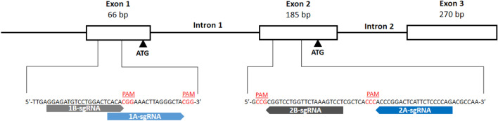
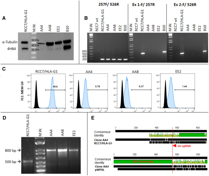
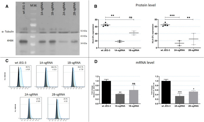
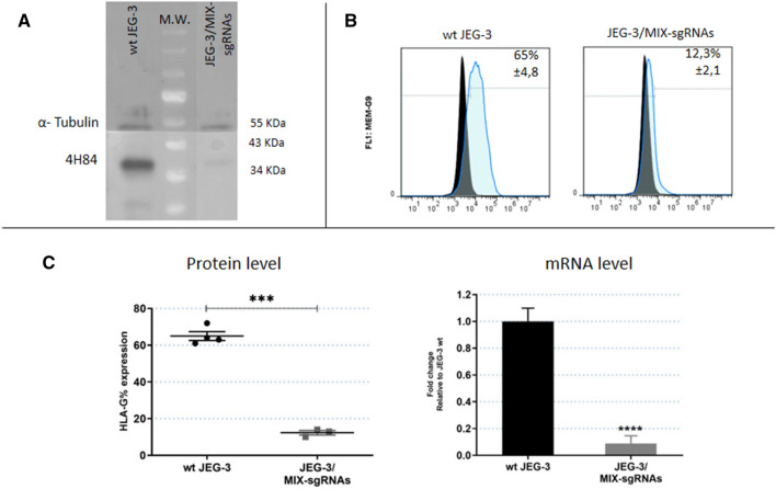
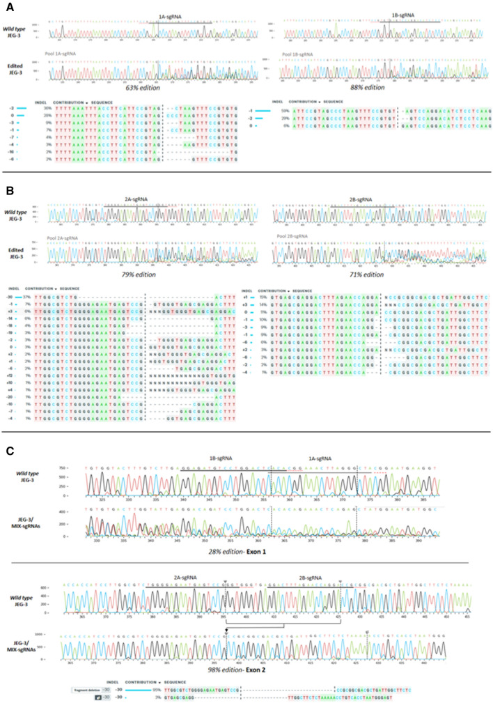
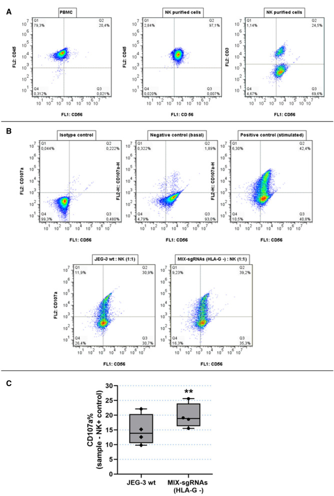
Cancer immunotherapies based mainly on the blockade of immune-checkpoint (IC) molecules by anti-IC antibodies offer new alternatives for treatment in oncological diseases. However, a considerable proportion of patients remain unresponsive to them. Hence, the development of novel clinical immunotherapeutic approaches and/or targets are crucial.W In this context, targeting the immune-checkpoint HLA-G/ILT2/ILT4 has caused great interest since it is abnormally expressed in several malignancies generating a tolerogenic microenvironment. Here, we used CRISPR/Cas9 gene editing to block the HLA-G expression in two tumor cell lines expressing HLA-G, including a renal cell carcinoma (RCC7) and a choriocarcinoma (JEG-3). Different sgRNA/Cas9 plasmids targeting HLA-G exon 1 and 2 were transfected in both cell lines. Downregulation of HLA-G was reached to different degrees, including complete silencing. Most importantly, HLA-G - cells triggered a higher in vitro response of immune cells with respect to HLA-G + wild type cells. Altogether, we demonstrated for the first time the HLA-G downregulation through gene editing. We propose this approach as a first step to develop novel clinical immunotherapeutic approaches in cancer.


Conflict of interest statement

The authors declare no competing interests.

Figures

Figure 1
Figure 1
Schematic representation of the first 3 exons of HLA-G gene and the 4 designed sgRNAs. The white boxes and lines represent exons and introns, respectively. The sequence below represents part of exon 1 and 2 containing Cas9/sgRNA target sites for 1A-, 1B-, 2A- and 2B-sgRNAs. Protospacer-adjacent motif (PAM) is labeled in red. Black arrows represent the two transcription start sites.
Figure 2
Figure 2
Analysis of RCC7/HLA-G1 edited cells by CRISPR/Cas9: (A) Western Blot analysis of HLA-G expression in 2A-sgRNA clonal cell lines (right). Wild type RCC7/HLA-G1 control is shown on the left. (B) RT-PCR to analyze mRNA expression. (C) Flow cytometry analysis of HLA-G protein expression. (D) Genomic DNA amplified by PCR. (E) Sequence alignment of wild type RCC7/HLA-G1 vs. clone AA4 and pWXPL-lentivirus vector. The red arrow represents the 2A-sgRNA target site and the red line shows the cut site of Cas9 protein. http://www.geneious.com.
Figure 3
Figure 3
Analysis of JEG-3 edited cells by CRISPR/Cas9 with each sgRNA (1A-, 1B-, 2A- and 2B) (A) Western Blot analysis of HLA-G expression in JEG-3 edited cells (right) and wild type JEG-3 (left). (B) Protein expression measured by flow cytometry. (C) Representative histograms of HLA-G measured by flow cytometry. Isotype control is shown in black. (D) The HLA-G mRNA expression measured by RT-qPCR. No significant: ns. Significant differences are shown with * (p < 0.05).
Figure 4
Figure 4
Analysis of JEG-3 edited cells by CRISPR/Cas9 with 4 sgRNAs simultaneously (named MIX-sgRNAs) (A) Western Blot analysis of HLA-G expression in JEG-3/MIX-sgRNAs cells (right) and wild type JEG-3 (left). (B) Representative histograms of HLA-G measured by flow cytometry. Isotype control is shown in black. (C) On the left is shown the protein expression measured by flow cytometry. On the right is shown the HLA-G mRNA expression measured by RT-qPCR. No significant: ns. Significant differences are shown with * (p < 0.05).
Figure 5
Figure 5
Sequencing analysis. Histograms with nucleotide sequence data of cell pools edited by CRISPR system. Different edited genotypes predicted by InDels analysis (https://www.ice.synthego.com/) and percentage of edition for each condition: (A) JEG-3 cell pools edited with 1A- and 1B-sgRNAs, in exon 1 region. (B) JEG-3 cell pools edited with 2A- and 2B-sgRNAs, in exon 2 region. (C) JEG-3 cell pools edited with all sgRNAs (1A, 1B, 2A and 2B, named JEG-3/MIX-sgRNAs), in exon 1 and 2 simultaneously.
Figure 6
Figure 6
Degranulation assay, functional analysis of HLA-G wt and HLA-G − JEG-3 cells. (A) Determination of NK cells purification. NK cells were stained with anti-CD45/PE, anti-CD56/BB515 and anti-CD3/PE and compared with PBMC. (B) As a representative assay, NKs were labelled with CD107a/PE and CD56-BB515. The conditions were the following: Isotype control (NK without Ab), negative control (NK basal degranulation), positive control (stimulated NK cells) and NKs co-cultured with wt JEG-3 or with JEG-3/MIX-sgRNAs (HLA-G −). (C) Box plots show the percentage of CD107a + NK cells when co-cultured with wt JEG-3 (HLA-G wt) or with JEG-3/MIX-sgRNAs (HLA-G −). Significant differences are shown with * (p < 0.05) (n = 4 experiments).

References

    1. Marin-Acevedo JA, et al. Next generation of immune checkpoint therapy in cancer: New developments and challenges. J. Hematol. Oncol. 2018;11:1–20. - PMC - PubMed
    1. Hahn AW, Gill DM, Pal SK, Agarwal N. The future of immune checkpoint cancer therapy after PD-1 and CTLA-4. Immunotherapy. 2017;9:681–692. - PubMed
    1. Weber J. Immune checkpoint proteins: A new therapeutic paradigm for cancerpreclinical background: CTLA-4 and PD-1 blockade. Semin. Oncol. 2010;37:430–439. - PubMed
    1. Collin M. Immune checkpoint inhibitors: A patent review (2010–2015) Expert Opin. Ther. Pat. 2016;26:555–564. - PubMed
    1. Hargadon KM, Johnson CE, Williams CJ. Immune checkpoint blockade therapy for cancer: An overview of FDA-approved immune checkpoint inhibitors. Int. Immunopharmacol. 2018;62:29–39. - PubMed

Substances