

Ethmoidal silent sinus syndrome after nasal swab test
- PMID: 34773478
- PMCID: PMC8589631
- DOI: 10.1007/s00234-021-02856-y
Ethmoidal silent sinus syndrome after nasal swab test
Abstract
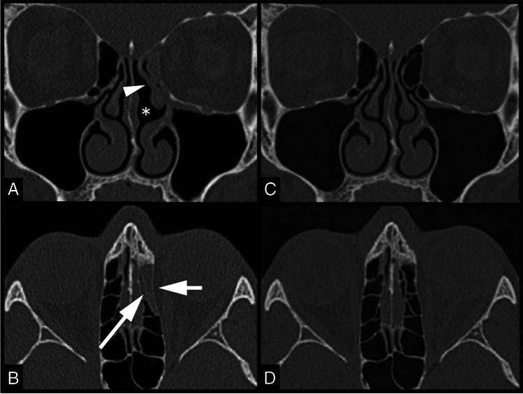
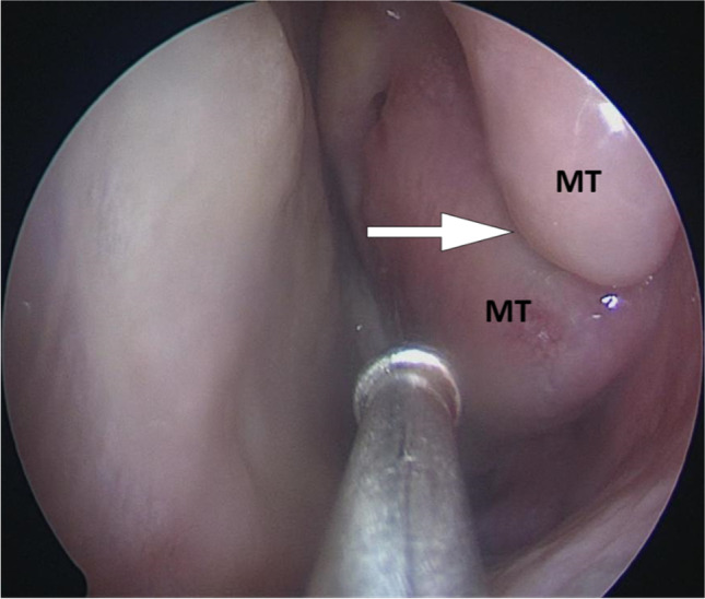
This study reported the case of a healthy male in his 40s who presented with a 3-month history of frontal headache and post-nasal drip, which did not improve with oral antibiotics. One month prior to the onset of the symptoms, he underwent a nasopharyngeal swab testing for SARS-CoV-2 (which yielded a negative result) for a history of malaise and cough. The patient claimed that the swab insertion into the nasal cavity was particularly painful on the left side. Sinus computed tomography (CT) scan showed deformity of the left middle nasal turbinate with occlusion of the osteomeatal complex, resulting in ethmoid silent sinus syndrome. This study makes a significant contribution to the literature because nasopharyngeal, midturbinate and anterior nasal swabs have been recommended as initial diagnostic specimen collection methods by the US Centers for Disease Control and Prevention (CDC) for the coronavirus disease 2019. These methods require introducing an instrument into the nasal cavity, potentially leading to adverse effects due to the delicate and complex nasal anatomy. However, complications related to swab testing for respiratory pathogens have not yet been fully discussed in the literature.
Keywords: COVID-19; Coronavirus; Nasopharyngeal swab; Silent sinus syndrome.
© 2021. The Author(s), under exclusive licence to Springer-Verlag GmbH Germany, part of Springer Nature.
Conflict of interest statement
The authors have no conflicts of interest to declare that are relevant to the content of this article.
Figures
References
-
- Centers for Disease Control and Prevention (2021) Interim guidelines for collecting, handling, and testing clinical specimens for COVID-19. https://www.cdc.gov/coronavirus/2019-ncov/lab/guidelines-clinical-specim.... Accessed 30 October 2021
-
- Fabbris C, Cestaro W, Menegaldo A, Spinato G, Frezza D, Vijendren A, Borsetto D, Boscolo-Rizzo P. Is oro/nasopharyngeal swab for SARS-CoV-2 detection a safe procedure? Complications observed among a case series of 4876 consecutive swabs. Am J Otolaryngol. 2021;42:102758. doi: 10.1016/j.amjoto.2020.102758. - DOI - PMC - PubMed
-
- Montgomery WW. Mucocele of the maxillary sinus causing enophthalmos. Eye Ear Nose Throat Mon. 1964;43:41–42. - PubMed
MeSH terms
LinkOut - more resources
Full Text Sources
Medical
Miscellaneous
