An official website of the United States government
The .gov means it’s official.
Federal government websites often end in .gov or .mil. Before
sharing sensitive information, make sure you’re on a federal
government site.
The site is secure.
The https:// ensures that you are connecting to the
official website and that any information you provide is encrypted
and transmitted securely.
1 Department of Physiology, Faculty of Medicine and Dentistry, University of Alberta, Edmonton, Alberta, Canada; Mazankowski Alberta Heart Institute, University of Alberta, Edmonton, Alberta, Canada.
2 Department of Physiology, Faculty of Medicine and Dentistry, University of Alberta, Edmonton, Alberta, Canada.
3 Faculty of Pharmacy and Pharmaceutical Sciences, University of Alberta, Edmonton, Alberta, Canada.
4 Faculty of Pharmacy and Pharmaceutical Sciences, University of Alberta, Edmonton, Alberta, Canada; Department of Pharmacology, Faculty of Medicine and Dentistry, University of Alberta, Edmonton, Alberta, Canada.
5 Mazankowski Alberta Heart Institute, University of Alberta, Edmonton, Alberta, Canada; Division of Cardiology, Department of Medicine, Faculty of Medicine and Dentistry, University of Alberta, Edmonton, Alberta, Canada. Electronic address: gavin.oudit@ualberta.ca.
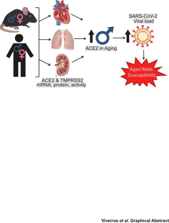
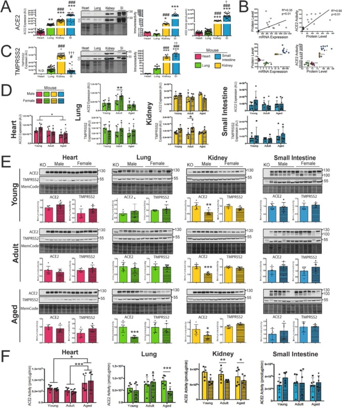
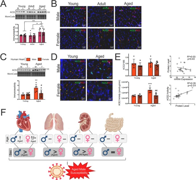
Aged males disproportionately succumb to increased COVID-19 severity, hospitalization, and mortality compared to females. Angiotensin-converting enzyme 2 (ACE2) and transmembrane protease, serine 2 (TMPRSS2) facilitate SARS-CoV-2 viral entry and may have sexually dimorphic regulation. As viral load dictates disease severity, we investigated the expression, protein levels, and activity of ACE2 and TMPRSS2. Our data reveal that aged males have elevated ACE2 in both mice and humans across organs. We report the first comparative study comprehensively investigating the impact of sex and age in murine and human levels of ACE2 and TMPRSS2, to begin to elucidate the sex bias in COVID-19 severity.
Assessment of ACE2 and TMPRSS2 across sex and aging. A. ACE2 mRNA expression…
Fig. 1
Assessment of ACE2 and TMPRSS2 across sex and aging. A. ACE2 mRNA expression (n = 36, biological replicates), protein levels (n = 11, biological replicates), and activity (n = 36, biological replicates) are positively correlated across all organs in male and female animals (B.). C. TMPRSS2 mRNA (n = 36, biological replicates) and protein levels (n = 11, biological replicates). Data from A-C was generated from C57BL6/J mice using a pooled analysis of males and females. Levels are compiled from all sexes and age groups for expression and activity; however, protein levels are compiled from representative and three replicate gels sampled from a subset of all age groups (Supplementary Fig. S1). All organs were run on the same gel for comparison. Immunoblots were over-exposed (O.E.) to visualize low levels in the lung and heart. Expression and activity data were run one time in the same plate for quantification. Data are represented as median with interquartile range (IQR) and were analyzed as independent samples with the Kruskal-Wallis test with pairwise comparison adjusted by Bonferroni correction; * indicates differences from the heart; # indicates differences from the lung; and † indicates differences from the kidney. *p < 0.05; **p < 0.01, ***p < 0.001 where the number of symbols indicates the level of significance. Analysis of mRNA expression (D.), protein levels (E.), and ACE2 activity (F.) of ACE2 and TMPRSS2 across sex and age in the heart (magenta), lung (green), kidney (yellow), and small intestine (SI) (blue) (n = 6, biological replicates/group). ACE2 global knockout mice were analyzed as a negative control (KO) for western blots. Raw images are provided in Supplementary Fig. S2-S3. Data are represented as the mean ± SEM and analyzed by two-way ANOVA with Tukey post hoc test (mRNA and activity), or unpaired student's t-test (protein levels). For all experiments, young animals (n = 12) were obtained from four litters and each litter was collected over two days. Adult and aged animals (n = 12 /group) were obtained from five and three litters respectively, and each litter was collected over two days. (For interpretation of the references to colour in this figure legend, the reader is referred to the web version of this article.)
Fig. 2
Sex- and age-differences in ACE2…
Fig. 2
Sex- and age-differences in ACE2 levels in mouse and human hearts. A. Representative…
Fig. 2
Sex- and age-differences in ACE2 levels in mouse and human hearts. A. Representative immunoblot of mouse hearts for ACE2 protein levels (n = 6 biological replicates/group). Three gels were compiled for quantification (Supplementary Fig. S4). Data are represented as the mean ± SEM and were analyzed by two-way ANOVA with Tukey post hoc test; *p < 0.05; **p < 0.01, ***p < 0.001. For all experiments, young animals (n = 12) were obtained from four litters and each litter was collected over two days. Adult and aged animals (n = 12 /group) were obtained from five and three litters respectively, and each litter was collected over two days. B. Immunofluorescence of ACE2 (green) and pericyte marker NG2 (red). Wheat Germ Agglutinin (WGA) (blue) was used to delineate the cell membrane. Qualitative images were captured from n = 2 biological replicates and n = 16 (8 images/animal) technical replicates for each age group. C. Representative immunoblot for ACE2 in human hearts (n = 6, biological replicates/group). Two gels were compiled for quantification (Supplementary Fig. S4). Quantification represents the combined result from two western blots. Data are represented as the mean ± SEM and were analyzed by two-way ANOVA with Tukey post hoc test. * indicates differences from young males, and # indicates differences between aged males and aged females; *p < 0.05; **p < 0.01, ***p < 0.001 where the number of symbols indicates the level of significance. D. Immunofluorescence of ACE2 (green) and NG2 (red). WGA (blue) was used to delineate the cell membrane. Qualitative images were captured from n = 2 biological replicates and n = 16 (8 images/donor) technical replicates for each age group. E. ACE2 protein levels and activity are positively correlated, but not mRNA expression. * indicates differences from young males, and # indicates differences between aged males and aged females. *p < 0.05; **p < 0.01, ***p < 0.001 where the number of symbols indicates the level of significance. F. Schematic figure showing the multi-organ impact of sex and aging on ACE2 levels as a potential contributor to the increased male susceptibility to severe COVID-19. (For interpretation of the references to colour in this figure legend, the reader is referred to the web version of this article.)
Viveiros A., Rasmuson J., Vu J., Mulvagh S.L., Yip C.Y.Y., Norris C.M., et al. Sex differences in COVID-19: candidate pathways, genetics of ACE2, and sex hormones. Am. J. Physiol. Heart Circ. Physiol. 2021;320(1):H296–H304.
-
PMC
-
PubMed
Gheblawi M., Wang K., Viveiros A., Nguyen Q., Zhong J.C., Turner A.J., et al. Angiotensin-converting enzyme 2: SARS-CoV-2 receptor and regulator of the renin-angiotensin system: celebrating the 20th anniversary of the discovery of ACE2. Circ. Res. 2020;126(10):1456–1474.
-
PMC
-
PubMed
Muus C., Luecken M.D., Eraslan G., Sikkema L., Waghray A., Heimberg G., et al. Single-cell meta-analysis of SARS-CoV-2 entry genes across tissues and demographics. Nat. Med. 2021;27(3):546–559.
-
PMC
-
PubMed
Shenoy S. SARS-CoV-2 (COVID-19), viral load and clinical outcomes; lessons learned one year into the pandemic: a systematic review. World J. Crit. Care Med. 2021;10(4):132–150.
-
PMC
-
PubMed
Stelzig K.E., Canepa-Escaro F., Schiliro M., Berdnikovs S., Prakash Y.S., Chiarella S.E. Estrogen regulates the expression of SARS-CoV-2 receptor ACE2 in differentiated airway epithelial cells. Am. J. Physiol. Lung Cell Mol. Physiol. 2020;318(6):L1280–L1281.
-
PMC
-
PubMed