. 2021 Dec:279:121246.
doi: 10.1016/j.biomaterials.2021.121246. Epub 2021 Nov 10.

3D bioprinting of a trachea-mimetic cellular construct of a clinically relevant size

Affiliations

3D bioprinting of a trachea-mimetic cellular construct of a clinically relevant size

Jeong Hun Park et al. Biomaterials. 2021 Dec.

Abstract

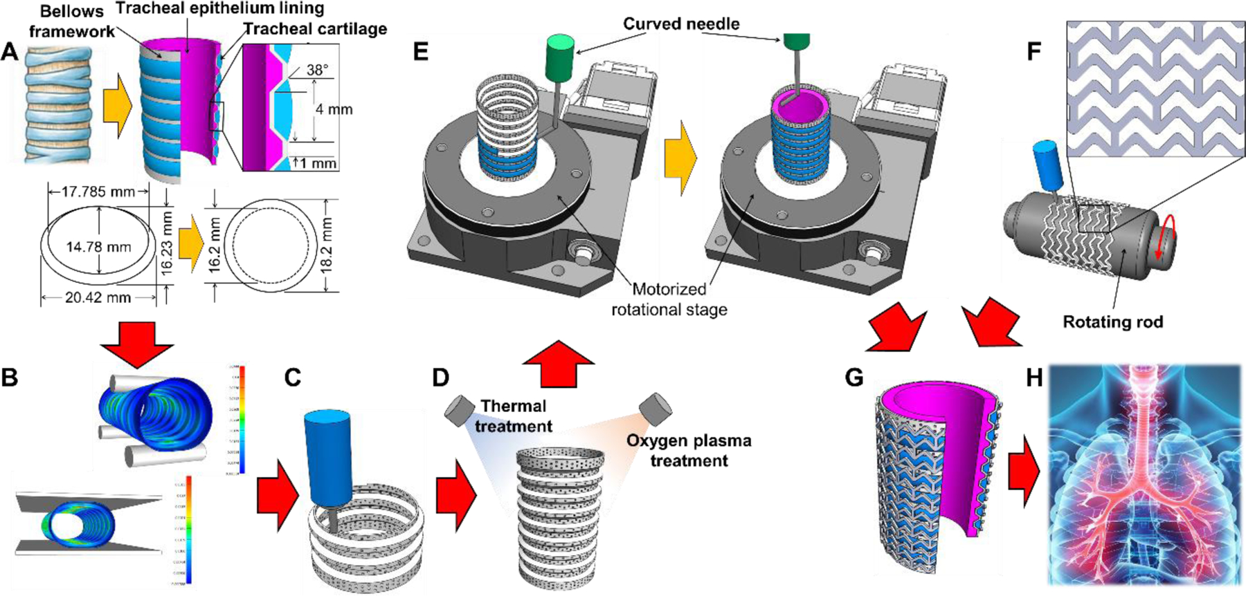
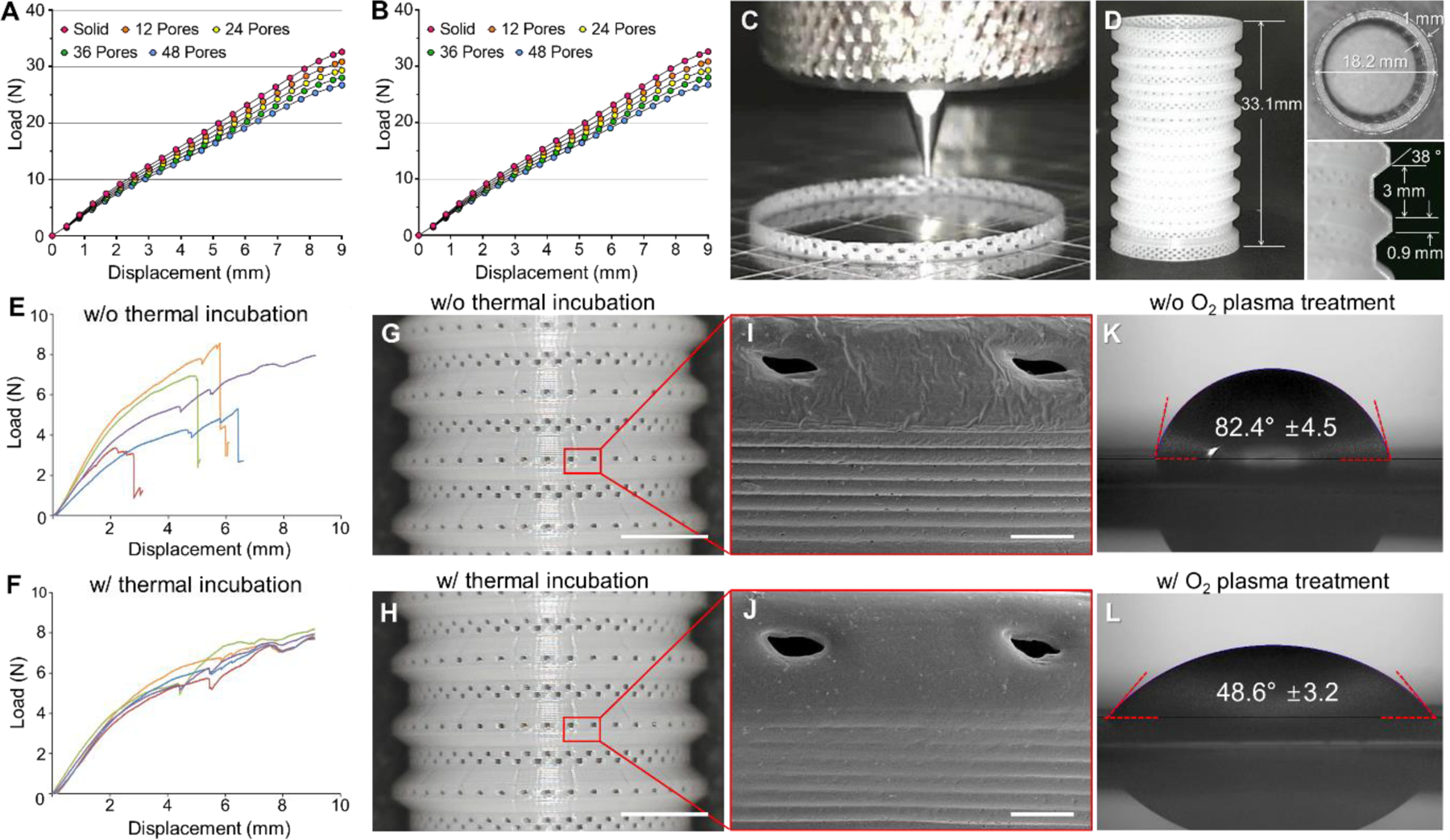
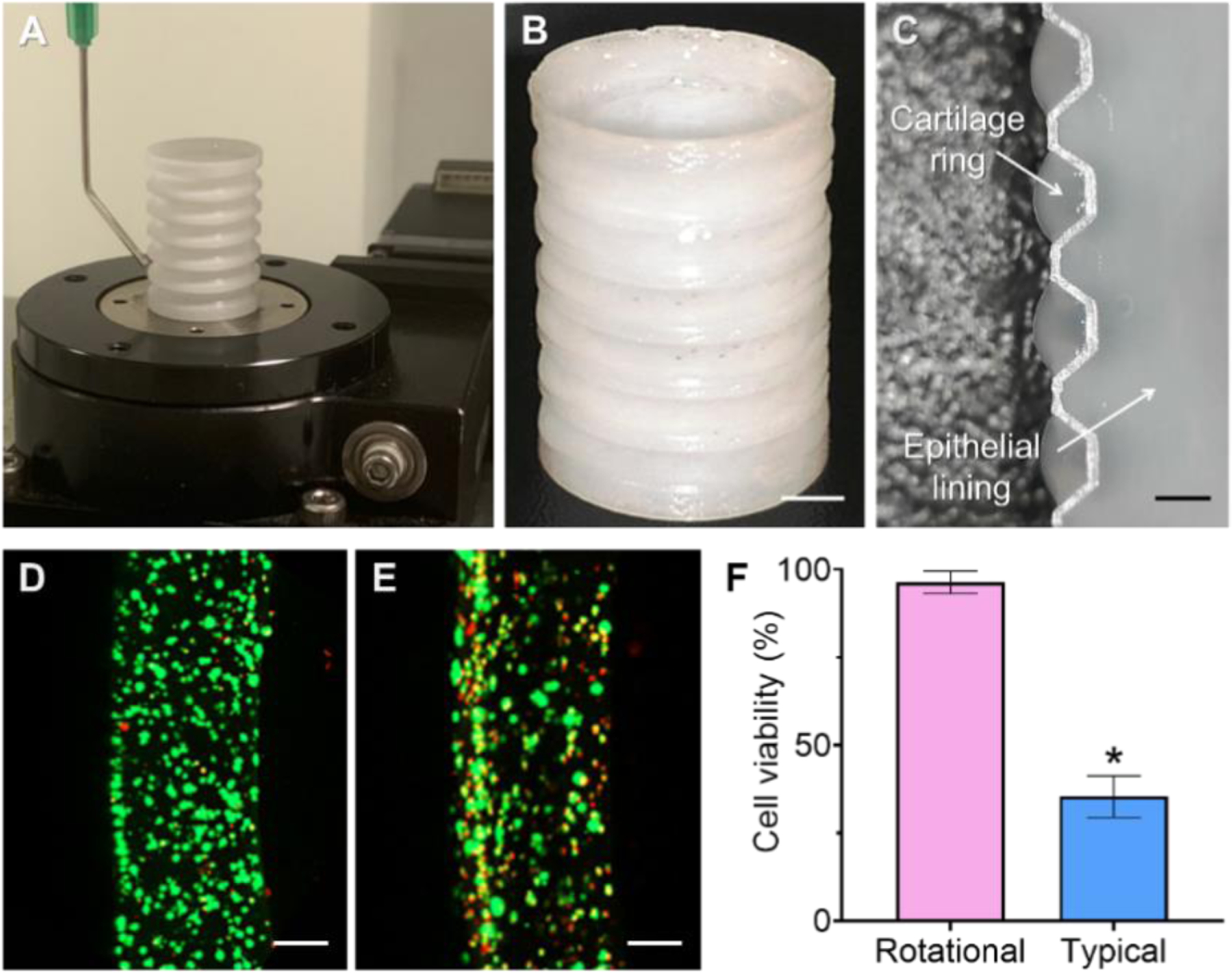
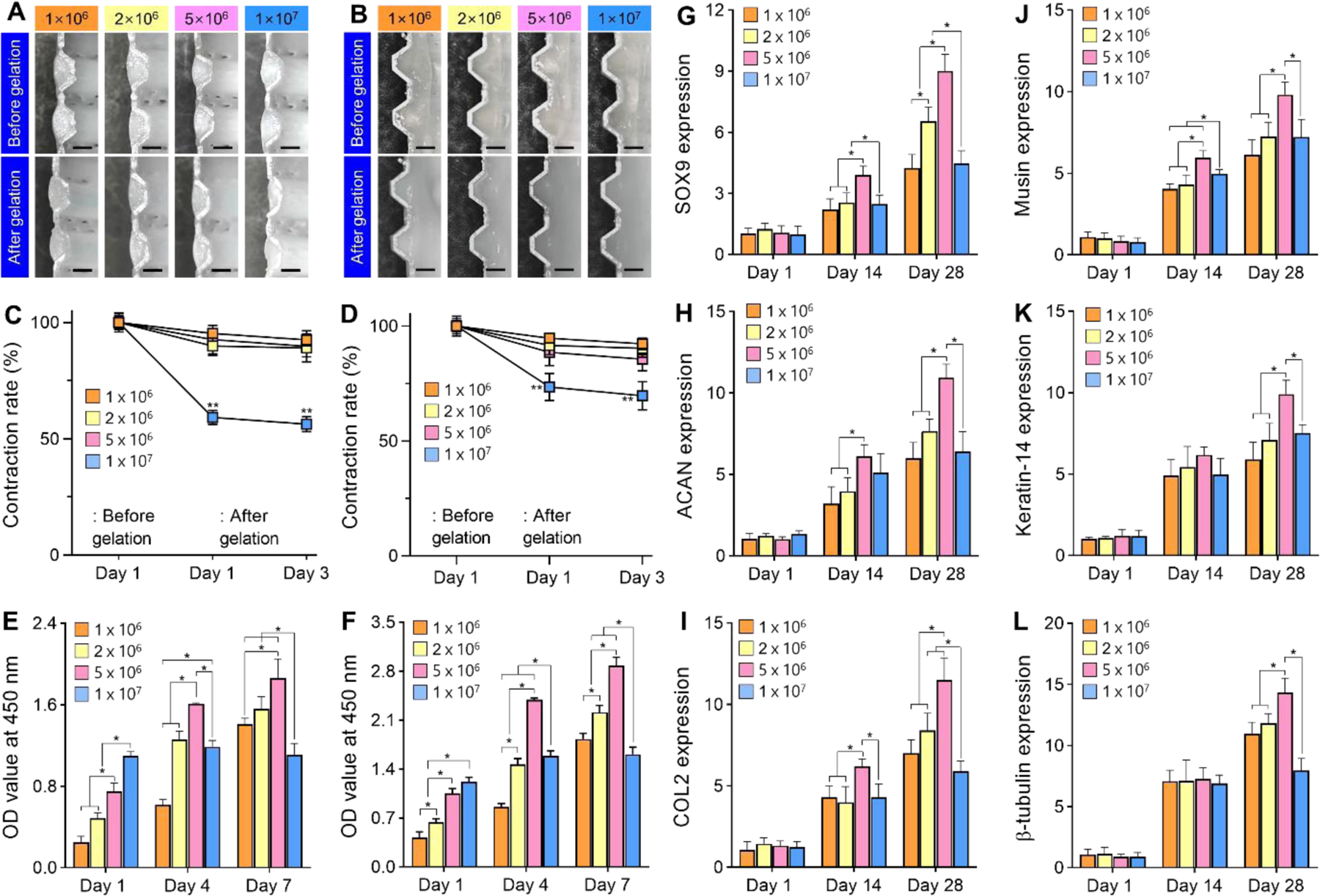
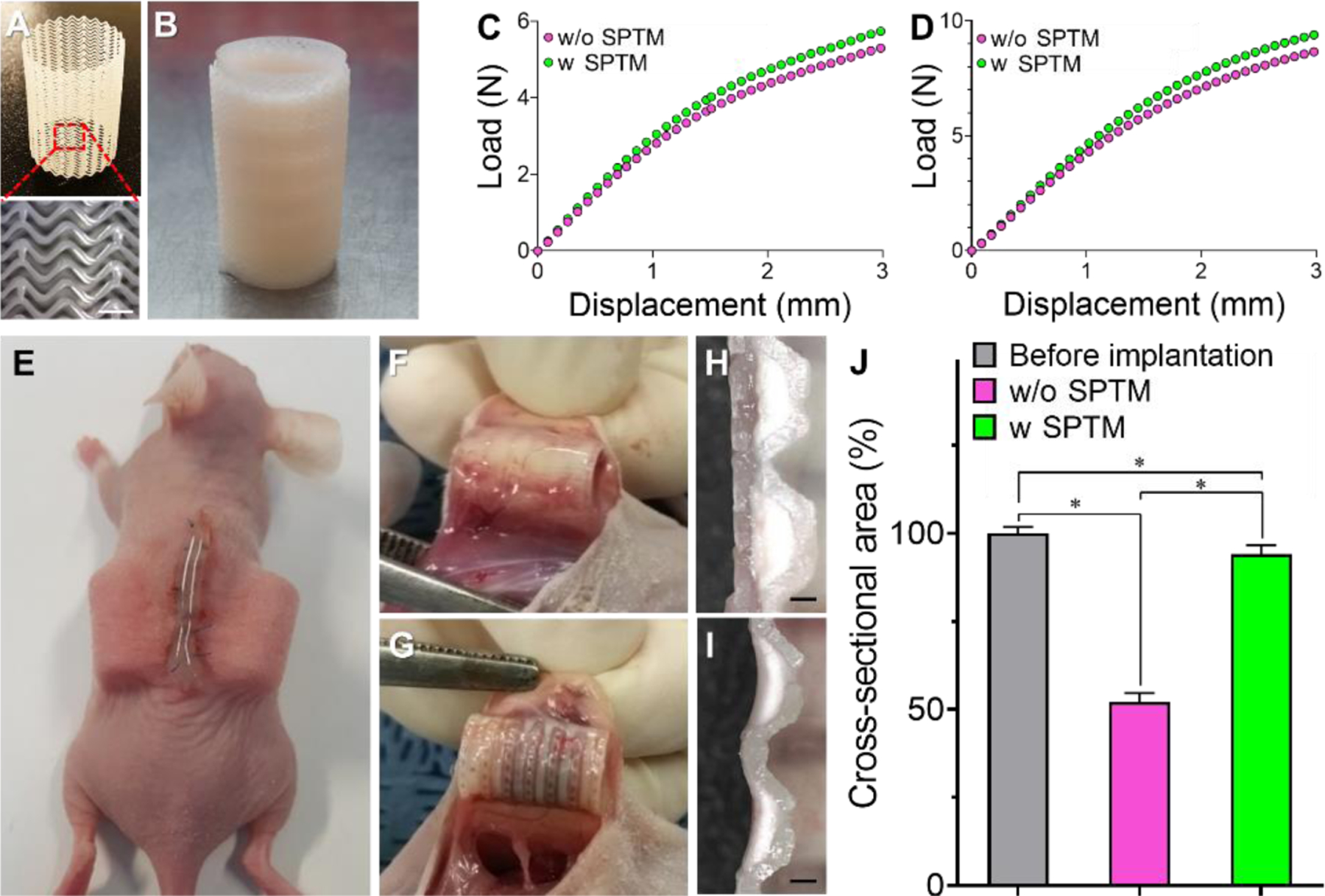
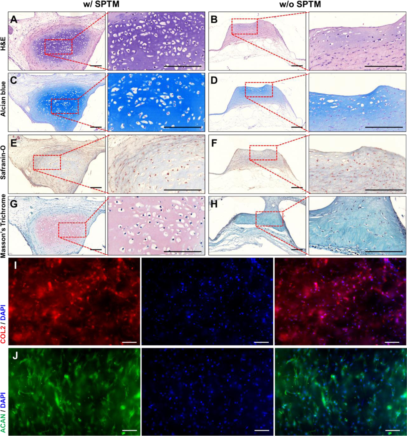
Despite notable advances in extrusion-based 3D bioprinting, it remains a challenge to create a clinically-sized cellular construct using extrusion-based 3D printing due to long printing times adversely affecting cell viability and functionality. Here, we present an advanced extrusion-based 3D bioprinting strategy composed of a two-step printing process to facilitate creation of a trachea-mimetic cellular construct of clinically relevant size. A porous bellows framework is first printed using typical extrusion-based 3D printing. Selective printing of cellular components, such as cartilage rings and epithelium lining, is then performed on the outer grooves and inner surface of the bellows framework by a rotational printing process. With this strategy, 3D bioprinting of a trachea-mimetic cellular construct of clinically relevant size is achieved in significantly less total printing time compared to a typical extrusion-based 3D bioprinting strategy which requires printing of an additional sacrificial material. Tracheal cartilage formation was successfully demonstrated in a nude mouse model through a subcutaneous implantation study of trachea-mimetic cellular constructs wrapped with a sinusoidal-patterned tubular mesh preventing rapid resorption of cartilage rings in vivo. This two-step 3D bioprinting for a trachea-mimetic cellular construct of clinically relevant size can provide a fundamental step towards clinical translation of 3D bioprinting based tracheal reconstruction.

Keywords: 3D bioprinting; Clinically relevant size; Trachea-mimetic cellular construct; Tracheal cartilage regeneration.


Conflict of interest statement

Declaration of competing interests

J.H.P., M.A., S.-H.P., J.-S.L., S.W.K., and D.-W.C. are inventors on a patent related to this work. The authors declare that they have no competing interests.

Declaration of interests

The authors declare that they have no known competing financial interests or personal relationships that could have appeared to influence the work reported in this paper.

Figures

Fig. 1.
Fig. 1.
Schematics of the advanced two-step extrusion-based 3D bioprinting of the trachea-mimetic cellular construct of clinically relevant size. Step 1: (A) Design of the trachea-mimetic cellular construct based on canine tracheal anatomy. (B) FEA determining the effect of pore distribution on the mechanical behavior of the bellows framework. (C) Creation of porous bellows framework using a typical extrusion-based 3D printing. (D) Thermal/oxygen plasma treatments on the printed bellows framework. Step 2: (E) Rotational printing of separate cartilage rings and epithelial lining on the outer grooves and on the luminal surface of the bellows framework. (F) Creation of a SPTM for prevention of rapid resorption of cartilage rings in vivo. (G) Wrapping the trachea-mimetic cellular construct with a SPTM. (H) Implantation of the trachea-mimetic cellular construct wrapped with a SPTM into the circumferential tracheal defect for simultaneous regeneration of tracheal cartilage and epithelium.
Fig. 2.
Fig. 2.
Mechanical behavior analysis and extrusion-based 3D bioprinting of bellows frameworks. FEA load-displacement curves of bellows framework having different pore distributions under (A) radial compression and (B) three-point bending. (C) Photograph of typical extrusion-based printing process of porous bellows framework. (D) Printing results of porous bellows framework of clinically relevant size. Load-displacement curves of porous bellows framework under three-point bending (E) before and (F) after thermal incubation. Photographs of the PCL bellows framework (G) before and (H) after thermal incubation. Scale bars, 5 mm. SEM images of the red box in each photograph of bellows framework (I) before and (J) after thermal incubation for pore architecture observation. Scale bars, 500 μm. Contact angle of water droplet on the outer surface of bellows framework (K) before oxygen plasma treatment and (L) after oxygen plasma treatment.
Fig. 3.
Fig. 3.
Rotational printing of separate cartilage rings and epithelial lining. (A) Photograph of rotational printing process. (B) Trachea-mimetic cellular construct after rotational printing. Scale bar, 5 mm. (C) Longitudinal cross-section of a trachea-mimetic cellular construct along with an interior surface. Scale bar, 1 mm. Radial cross-section of cartilage ring stained for live and dead cells from (D) rotational printing strategy and (E) typical printing strategy. Scale bars, 200 μm. (F) Cell viability according to different printing strategies. *p<0.01.
Fig. 4.
Fig. 4.
Cell density effect on the biological performance of cartilage rings and epithelial lining. Longitudinal cross-sections of (A) cartilage rings and (B) epithelial lining of tracheal construct with different cell densities before and after gelation. Scale bars, 10 mm. Volume contraction rate on (C) cartilage rings and (D) epithelial lining over time in culture for 3 days. Proliferation of (C) hNCs in cartilage rings and (F) hNTSCs in epithelial lining with different cell densities for 7 days of culture. Gene expression levels of cellular tracheal constructs with different cell densities for 28 days of culture using chondrogenic representative markers of (G) SOX9, (H) ACAN, and (I) COL2 and epithelial markers of (J) Musin, (K) Keratin-14, and (L) β-tubulin. *p<0.01. **p<0.01 when compared with the other groups.
Fig. 5.
Fig. 5.
Prevention of cartilage rings resorption in vivo. (A) Photograph of SPTM. (B) Photograph of a trachea-mimetic cellular construct wrapping with SPTM. Scale bar, 1 mm. Load-displacement curves of trachea-mimetic constructs with or without SPTM under (C) three-point bending and (D) radial compression. (E) Subcutaneous implantation of trachea-mimetic constructs with or without SPTM wrapping. Retrieval of trachea-mimetic tracheal construct (F) with and (G) without SPTM wrapping after 8 weeks of implantation. Longitudinal cross-section of cartilage rings of retrieved trachea-mimetic construct (H) with and (I) without wrapping of SPTM at 8 weeks after implantation. Scale bars, 1 mm. (J) Cross-sectional area of cartilage rings from trachea-mimetic constructs before implantation and retrieved trachea-mimetic constructs after 8 weeks of implantation. *p<0.01.
Fig. 6.
Fig. 6.
Evaluation of cartilage formation of trachea-mimetic cellular construct in vivo. (A, B) The H&E, (C, D) Alcian Blue, (E, F) Safranin-O, and (G, H) Masson’s Trichrome staining result of longitudinal cross-section of cartilage rings from retrieved cellular constructs with (left) and without (right) wrapping of SPTM after 8 weeks of implantation. Scale bars, 500 μm. The immunofluorescence image of (I) COL2 and (J) ACAN in the retrieved trachea-mimetic cellular tracheal construct with wrapping of SPTM after 8 weeks of implantation. Scale bars, 200 μm.

References

    1. Luo X, Liu Y, Zhang Z, Tao R, Liu Y, He A, Yin Z, Li D, Zhang W, Liu W, Long-term functional reconstruction of segmental tracheal defect by pedicled tissue-engineered trachea in rabbits, Biomaterials 34(13) (2013) 3336–3344. - PubMed
    1. Dhasmana A, Singh A, Rawal S, Biomedical grafts for tracheal tissue repairing and regeneration “Tracheal tissue engineering: an overview”, Journal of Tissue Engineering and Regenerative Medicine (2020). - PubMed
    1. Bae S-W, Lee K-W, Park J-H, Lee J, Jung C-R, Yu J, Kim H-Y, Kim D-H, 3D bioprinted artificial trachea with epithelial cells and chondrogenic-differentiated bone marrow-derived mesenchymal stem cells, International journal of molecular sciences 19(6) (2018) 1624. - PMC - PubMed
    1. Ke D, Yi H, Est-Witte S, George S, Kengla C, Lee SJ, Atala A, Murphy SV, Bioprinted trachea constructs with patient-matched design, mechanical and biological properties, Biofabrication 12(1) (2019) 015022. - PubMed
    1. Kim SH, Yeon YK, Lee JM, Chao JR, Lee YJ, Seo YB, Sultan MT, Lee OJ, Lee JS, S.-i. Yoon, Precisely printable and biocompatible silk fibroin bioink for digital light processing 3D printing, Nature communications 9(1) (2018) 1–14. - PMC - PubMed

Publication types