. 2022 Sep;92(3):712-720.
doi: 10.1038/s41390-021-01832-9. Epub 2021 Nov 13.

Dose- and sex-dependent effects of phlebotomy-induced anemia on the neonatal mouse hippocampal transcriptome

Affiliations

Dose- and sex-dependent effects of phlebotomy-induced anemia on the neonatal mouse hippocampal transcriptome

Garima Singh et al. Pediatr Res. 2022 Sep.

Abstract

Background: Phlebotomy-induced anemia (PIA) is universal and variable in degree among preterm infants and may contribute to neurodevelopmental risk. In mice, PIA causes brain tissue hypoxia, iron deficiency, and long-term sex-dependent neurobehavioral abnormalities. The neuroregulatory molecular pathways disrupted by PIA underlying these effects are unknown.

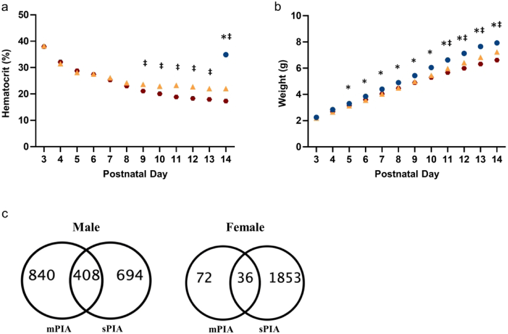
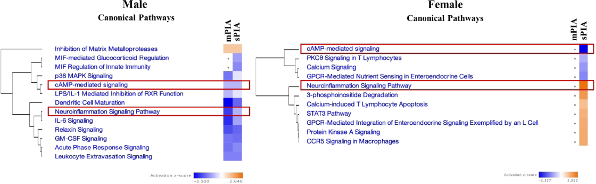
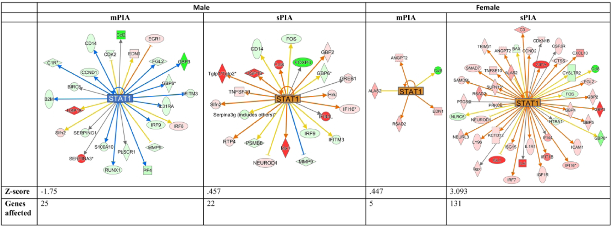
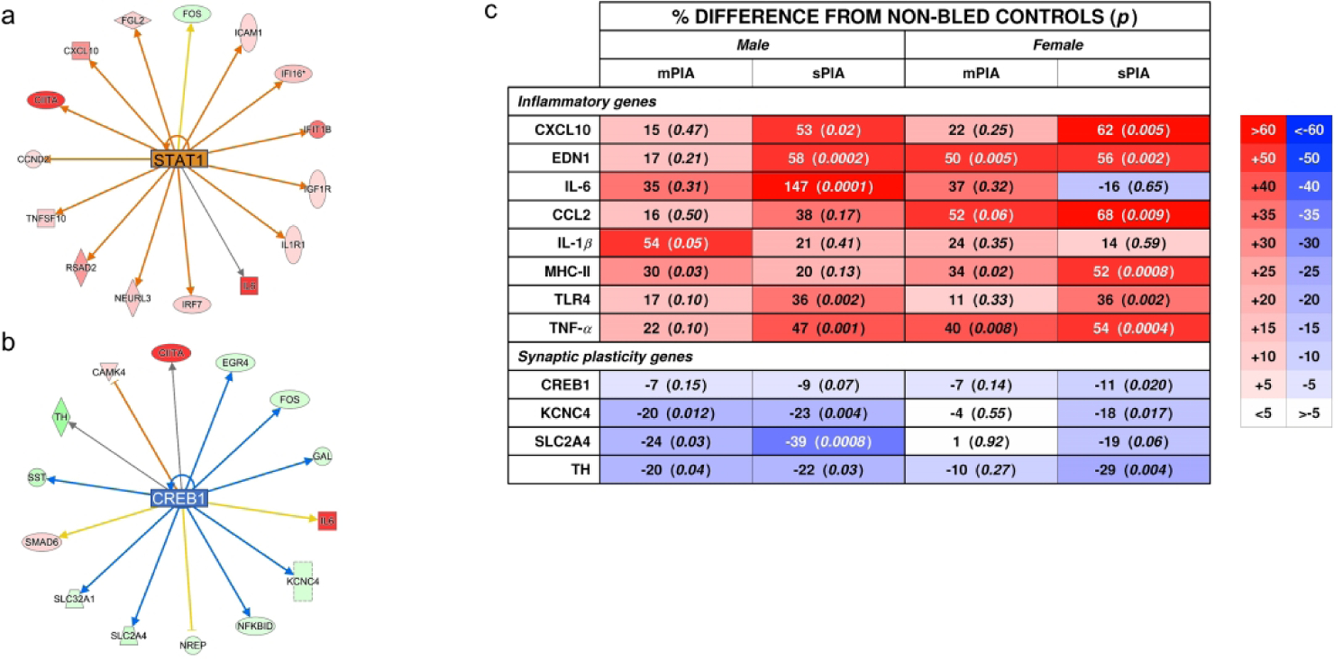
Methods: Male and female pups were phlebotomized daily from postnatal day (P)3-P14 via facial venipuncture to target hematocrits of 25% (moderate, mPIA) and 18% (severe, sPIA). P14 hippocampal RNA from non-bled control and PIA mice was sequenced by next-generation sequencing to identify differentially expressed genes (DEGs) that were analyzed using Ingenuity Pathway Analysis.

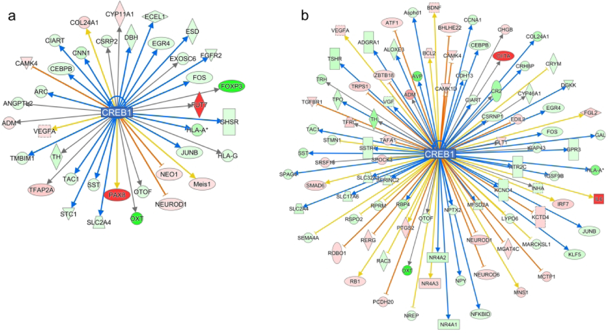
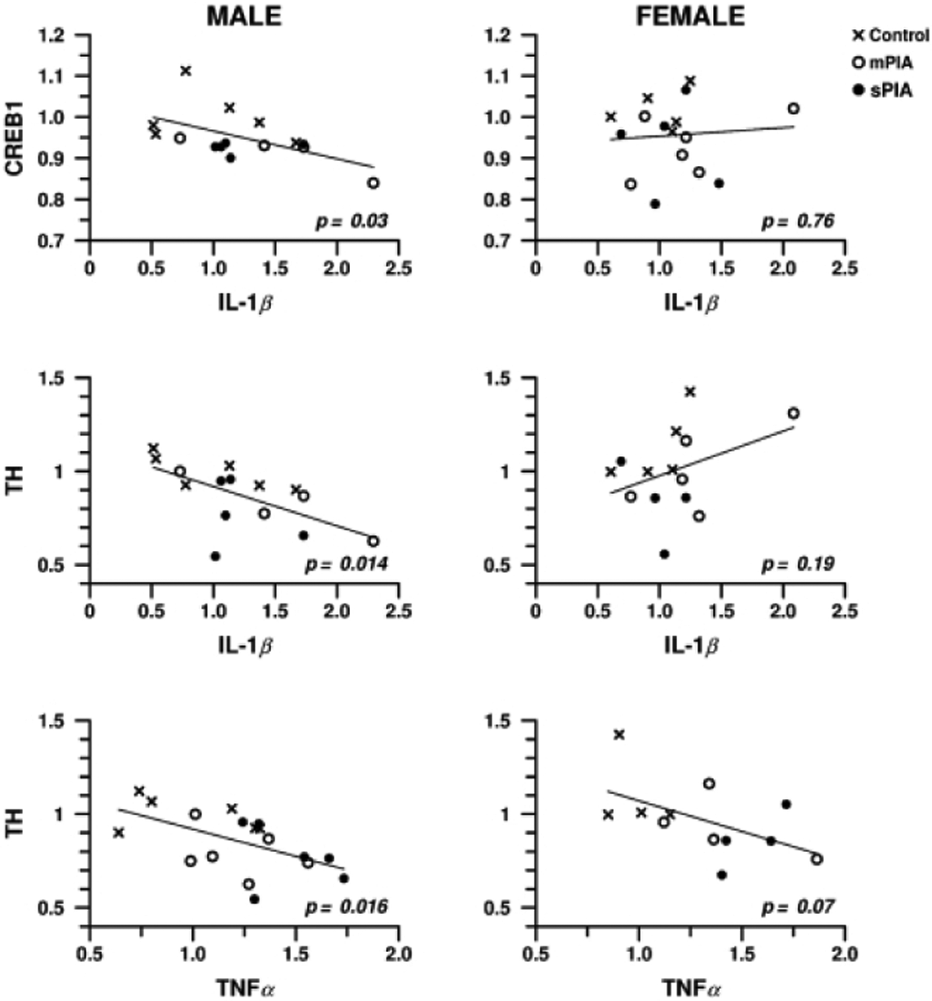
Results: mPIA females showed the least DEGs (0.5% of >22,000 genes) whereas sPIA females had the most (8.6%), indicating a dose-dependent effect. mPIA and sPIA males showed similar changes in gene expression (5.3% and 4.7%, respectively), indicating a threshold effect at mPIA. The pattern of altered genes induced by PIA indicates sex-specific and anemia-dose-dependent effects with increased pro-inflammation in females and decreased neurodevelopment in males.

Conclusion: These gene-expression changes may underlie the reduced recognition memory function in male and abnormal social-cognitive behavior in female adult mice following neonatal PIA. These results parallel clinical studies demonstrating sex-specific behavioral outcomes as a function of neonatal anemia.

Impact: Phlebotomy-induced anemia (PIA) in neonatal mice results in an altered hippocampal transcriptome and the severity of changes are dependent upon degree of anemia and sex of neonatal mice. The reported findings provide context to the sex-specific outcomes that have been reported in transfusion threshold clinical trials of preterm infants and therefore may inform treatment strategies that may be based on sex. These data advance the field by showing that consequences of PIA may be based in sex-specific transcriptomic alterations. Such changes may also result from other causes of neonatal anemia that also affect term infants.


Conflict of interest statement

Disclosure statement: The authors declare no financial competing interests.

Figures

Figure 1:
Figure 1:. Hematocrit and Weight trajectories of mPIA and sPIA animals and distribution of novel and common dysregulated genes.
(A) Daily hematocrit values for each group. (B) Daily weight values for each group. Blue circles = NBC; Yellow triangles = mPIA; Red circles = sPIA. *p< 0.05 NBC vs mPIA and sPIA; ‡p<0.05 mPIA vs sPIA. Graphs represent males and females combined (n= 64–92). (C) mPIA and sPIA males experienced a similar degree of dysregulation with mPIA resulting in dysregulation of 840 genes unique to the mPIA phenotype and 694 genes unique to the sPIA phenotype. 408 genes were common to both cohorts. Females experienced relatively minor gene dysregulation at mPIA but were severely affects at sPIA with 1853 unique dysregulated genes.
Figure 2:
Figure 2:. Comparison analysis of dysregulated canonical pathways in PIA males and females.
(A) Males showed similar downregulation of pathways regardless of degree of anemia whereas females (B) showed dysregulation following sPIA only. Both sexes showed dysregulation of neuroinflammatory and cAMP-mediated signaling; directionality was sex-dependent for neuroinflammatory signaling, downregulation predicted for males and upregulation predicted for females.
Figure 3:
Figure 3:. STAT1 dysregulation across PIA severity in males and females.
sPIA females are predicted to have a significant upregulation of the STAT1 pathway. sPIA and mPIA males showed a similar dysregulation but opposite directionality. Red- and green-filled shapes indicate up- and downregulation of genes, respectively, with brighter color indicating larger differences. Orange lines indicate activation; blue lines indicate inhibition; yellow lines indicate findings inconsistent with the state of downstream activity; gray lines indicate that the effect is not predicted.
Figure 4:
Figure 4:. Inflammatory and synaptic plasticity genes affected by PIA.
Selected genes from the (A) STAT1 pathway and (B) CREB1 pathway were selected for confirmatory qPCR analyses along with other common inflammatory genes. Percentage difference from NBCs and p values are summarized in panel (C). Color intensity increases for increased difference. There was increased dysregulation after 25%, indicating a threshold response for males. Females showed a dose-dependent response to hematocrit for most genes at 18%.
Figure 5:
Figure 5:. CREB1 dysregulation in sPIA males and females.
CREB1 expression is predicted to be downregulated in both sPIA males (z score −3.38) and females (z-score −3.82) with more downstream genes being affected in females (36 v 93 genes). Red- and green-filled shapes indicate up- and downregulation of genes, respectively, with brighter color indicating larger differences. Orange lines indicate activation; blue lines indicate inhibition; yellow lines indicate findings inconsistent with the state of downstream activity; gray lines indicate that the effect is not predicted.
Figure 6:
Figure 6:. Regression analysis of synaptic plasticity genes as a function of neuroinflammation genes.
A negative correlation was observed for CREB1 as a function of IL-1β and TH as a function of Tnf-α and IL-1β in males. Females did not show a significant correlation.

References

    1. Bouyssi-Kobar M, et al. Third Trimester Brain Growth in Preterm Infants Compared With In Utero Healthy Fetuses. Pediatrics. 2016;138:e20161640. - PMC - PubMed
    1. Ajayi-Obe M, Saeed N, Cowan FM, Rutherford MA, Edwards AD. Reduced development of cerebral cortex in extremely preterm infants. Lancet. 2000;356:1162–1163. - PubMed
    1. Smyser CD, et al. Longitudinal analysis of neural network development in preterm infants. Cereb Cortex. 2010;20:2852–2862. - PMC - PubMed
    1. Kuzawa CW. Adipose tissue in human infancy and childhood: an evolutionary perspective. Am J Phys Anthropol. 1998;Suppl 27:177–209. - PubMed
    1. Bastian TW, von Hohenberg WC, Mickelson DJ, Lanier LM, Georgieff MK. Iron Deficiency Impairs Developing Hippocampal Neuron Gene Expression, Energy Metabolism, and Dendrite Complexity. Dev Neurosci. 2016;38:264–276. - PMC - PubMed

Publication types