. 2021 Oct 27:12:729367.
doi: 10.3389/fphar.2021.729367. eCollection 2021.

ROS/JNK/C-Jun Pathway is Involved in Chaetocin Induced Colorectal Cancer Cells Apoptosis and Macrophage Phagocytosis Enhancement

Affiliations

ROS/JNK/C-Jun Pathway is Involved in Chaetocin Induced Colorectal Cancer Cells Apoptosis and Macrophage Phagocytosis Enhancement

Huihui Wang et al. Front Pharmacol. .

Abstract

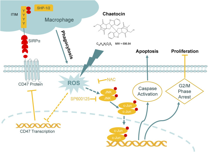
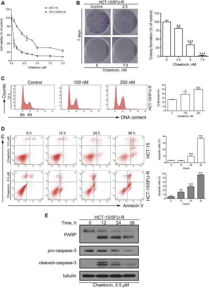
There is an urgent need for novel agents for colorectal cancer (CRC) due to the increasing number of cases and drug-resistance related to current treatments. In this study, we aim to uncover the potential of chaetocin, a natural product, as a chemotherapeutic for CRC treatment. We showed that, regardless of 5-FU-resistance, chaetocin induced proliferation inhibition by causing G2/M phase arrest and caspase-dependent apoptosis in CRC cells. Mechanically, our results indicated that chaetocin could induce reactive oxygen species (ROS) accumulation and activate c-Jun N-terminal kinase (JNK)/c-Jun pathway in CRC cells. This was confirmed by which the JNK inhibitor SP600125 partially rescued CRC cells from chaetocin induced apoptosis and the ROS scavenger N-acetyl-L-cysteine (NAC) reversed both the chaetocin induced apoptosis and the JNK/c-Jun pathway activation. Additionally, this study indicated that chaetocin could down-regulate the expression of CD47 at both mRNA and protein levels, and enhance macrophages phagocytosis of CRC cells. Chaetocin also inhibited tumor growth in CRC xenograft models. In all, our study reveals that chaetocin induces CRC cell apoptosis, irrelevant to 5-FU sensitivity, by causing ROS accumulation and activating JNK/c-Jun, and enhances macrophages phagocytosis, which suggests chaetocin as a candidate for CRC chemotherapy.

Keywords: CD47; JNK/c-Jun pathway; ROS; apoptosis; chaetocin; colorectal cancer.


Conflict of interest statement

The authors declare that the research was conducted in the absence of any commercial or financial relationships that could be construed as a potential conflict of interest.

Figures

Graph abstract
Graph abstract
Schematic representation of the mechanism of the action of chaetocin in CRC cells.
FIGURE 1
FIGURE 1
Chaetocin Inhibits cell proliferation and causes G2/M phase arrest. (A) Followed by treated with chaetocin at increasing concentrations for 24 h, CCK8 assay was used to measure the viability of CRC cells.(B) Real-time cell analysis results on the proliferation of HCT116, LS174T and LIM1215 cells. (C) After treated with chaetocin for indicated days, number of new colonies formed were counted. (D) Cell cycle distribution analysis on CRC cell lines after 12 h chaetocin treatment at various concentrations detected by PI-stained flow cytometry. For (C) and (D), results were shown as mean ± SD of three independent experiments. *p < 0.05, **p < 0.01, ***p < 0.001, versus control group.
FIGURE 2
FIGURE 2
Chaetocin induces caspase-dependent apoptosis in CRC cells. (A) Flow cytometry results with Annexin V-FITC/PI staining on apoptotic population of HCT116, LS174T and LIM1215 after chaetocin treatment. (B) Western blot analysis of PARP, caspase-3, capase-9 and caspase-8 expression levels in HCT116, LS174T and LIM1215 cells. (C) Flow cytometry results with JC-1 staining on three CRC cell lines. (D) Western blot results to evaluate the protein level of BCL-2, BCL-XL, MCL-1 and XIAP in CRC cell lines. (E) A 2 h z-VAD-fmk pretreatment could rescue some HCT116 and LS174T cells from apoptosis induced by chaetocin. For (A), (C) and (E), results were shown as mean ± SD of three independent experiments. *p < 0.05, **p < 0.01, ***p < 0.001. For (A) and (C), comparison was against 0 h group.
FIGURE 3
FIGURE 3
Chaetocin-induced ROS accumulation is required for cell apoptosis. (A) ROS levels of HCT116 and LS174T cells were verified using flow cytometry, after DCFH-DA labeling and chaetocin treatments. HCT116 and LS174T cells were pretreated with NAC for 2 h and then cotreated with chaetocin for another indicated times. (B) Flow cytometry analysis on apoptotic CRC cells. (C) Western blot of PARP and caspase-3 protein level. For (A) and (B), results were shown as mean ± SD of three independent experiments. *p < 0.05, **p < 0.01, ***p < 0.001, versus control group.
FIGURE 4
FIGURE 4
The involvement of ROS/JNK/c-Jun in ROS-mediated cell apoptosis. (A) The protein level of JNK and c-Jun in HCT116 and LS174T cells after chaetocin treatment by western blot. CRC cells were pretreated with SP600125 for 2 h before chaetocin treatment for indicated hours. (B) Cell apoptosis was measured by flow cytometry. Results were shown as mean ± SD of three independent experiments. **p < 0.01, ***p < 0.001. (C) Expression of PARP and caspase-3 was analyzed by western blot. (D) The expression levels of JNK and c-Jun after NAC pretreatment for 2 h and then cotreatment of chaetocin for 12 h in HCT116 and LS174T cells were analyzed by western blot.
FIGURE 5
FIGURE 5
Chaetocin overcomes 5-FU resistance in CRC cells. (A) The CCK8 cell viability assay on 5-FU resistant and sensitive HCT-15 cell lines. (B) After 7 days chaetocin treatment, colony formation result of HCT-15/5FU-R cells. (C) Cell cycle distribution results of HCT-15/5FU-R cells after 12 h chaetocin treatment at indicated concentration were analyzed by flow cytometry with PI staining. (D) Apoptotic population analysis by flow cytometry with Annexin V-FITC/PI staining of HCT-15 and HCT-15/5FU-R cells, after 0.5 μM chaetocin. (E) Western blot results on PARP and caspase-3 expression level in HCT-15/5FU-R cells. For a, (B), (C) and (D), results were shown as mean ± SD of three independent experiments. *p < 0.05, **p < 0.01, ***p < 0.001, versus control group.
FIGURE 6
FIGURE 6
Chaetocin-induced apoptosis in HCT-15/5FU-R cells is associated with the ROS/JNK/c-Jun pathway. (A) The ROS levels of HCT-15/5FU-R cells. Results were shown as mean ± SD of three independent experiments. ***p < 0.001, versus control group. (B) HCT-15/5FU-R cells apoptosis induced by chaetocin were rescued by 2 h of NAC pretreatment (***p < 0.001). (C) The expression level JNK and c-Jun protein in 5-FU-resistant CRC cells by western blot after chaetocin treatment. (D) HCT-15/5FU-R cells were cotreated with chaetocin for 24 h after 2 h SP600125 preincubation. Flow cytometry were used analyze the rescued population (***p < 0.001). (E) After pretreated with SP600125 for 2 h and then cotreated with chaetocin for another 6 h, the expression of JNK and c-Jun in HCT-15/5FU-R cells were analyzed by western blot. (F) The expression levels of JNK and c-Jun in HCT-15/5FU-R cells were evaluated by western blot after 2 h of NAC pretreatment followed by chaetocin cotreatment for 12 h.
FIGURE 7
FIGURE 7
Chaetocin decreases the expression of CD47 in CRC cells and enhances the phagocytosis of macrophages. (A) The expression of CD47 at mRNA levels was detected by qPCR after HCT116 and LS174T cells treated with chaetocin (0.5 μM chaetocin for HCT116 cells and 1 μM chaetocin for LS174T cells) for the indicated times. *p < 0.05, ***p < 0.001. (B) The expression of total CD47 protein was analyzed by western blot after chaetocin treatment. (C) The expression of membrane CD47 protein was detected by flow cytometry after HCT116 and LS174T were treated as described (*p < 0.05, **p < 0.01). (D) HCT116 cells were pretreated with 0.5 μM chaetocin for 24 h and then co-cultured with macrophages for another 24 h, the phagocytosis of HCT116 cells by macrophages was detected by flow cytometry. *p < 0.05. HCT116 cells were pretreated with 1 mM NAC for 2 h and then co-treated with 0.5 μM chaetocin for another 12 h (E) The mRNA level of CD47 was detected by qPCR. *p < 0.05, ***p < 0.001. (F) The expression of total CD47 protein was detected by western blot. SP600125 rescue analysis was done as previously mentioned. (G) mRNA expression of CD47 detection by qPCR (***p < 0.001). (H) The amount of CD47 protein expressed was evaluated by western blot.
FIGURE 8
FIGURE 8
Chaetocin inhibits the growth of CRC cell xenografts. Nude mice subcutaneously inoculated with HCT116 cells were subjected to chaetocin (0.5 mg/kg/day) treatment for 18 days, control group was treated with vehicle. (A) Tumor growth curves were recorded every other day.(B) and (C) The image of tumor tissue at the end of the experiment. (D) The protein levels of pro-caspase-3 and JNK in tumor tissues were detected by western blot. For (A) and (C), results were shown as mean ± SD of six mice in each group. *p < 0.05, versus vehicle group.

References

    1. Aggarwal V., Tuli H. S., Varol A., Thakral F., Yerer M. B., Sak K., et al. (2019). Role of Reactive Oxygen Species in Cancer Progression: Molecular Mechanisms and Recent Advancements. Biomolecules 9. 10.3390/biom9110735 - DOI - PMC - PubMed
    1. Biagi J. J., Raphael M. J., Mackillop W. J., Kong W., King W. D., Booth C. M. (2011). Association between Time to Initiation of Adjuvant Chemotherapy and Survival in Colorectal Cancer: a Systematic Review and Meta-Analysis. JAMA 305, 2335–2342. 10.1001/jama.2011.749 - DOI - PubMed
    1. Burgess T. L., Amason J. D., Rubin J. S., Duveau D. Y., Lamy L., Roberts D. D., et al. (2020). A Homogeneous SIRPα-CD47 Cell-Based, Ligand-Binding Assay: Utility for Small Molecule Drug Development in Immuno-Oncology. PLoS One 15, e0226661. 10.1371/journal.pone.0226661 - DOI - PMC - PubMed
    1. Capece D., Verzella D., Di Francesco B., Alesse E., Franzoso G., Zazzeroni F. (2019). NF-kappaB and Mitochondria Cross Paths in Cancer: Mitochondrial Metabolism and beyond. Semin. Cel Dev Biol 98, 118–128. 10.1016/j.semcdb.2019.05.021 - DOI - PubMed
    1. Castro-Caldas M., Carvalho A. N., Rodrigues E., Henderson C., Wolf C. R., Gama M. J. (2012). Glutathione S-Transferase Pi Mediates MPTP-Induced C-Jun N-Terminal Kinase Activation in the Nigrostriatal Pathway. Mol. Neurobiol. 45, 466–477. 10.1007/s12035-012-8266-9 - DOI - PubMed