Review
. 2021 Oct 27:12:748102.
doi: 10.3389/fphar.2021.748102. eCollection 2021.

Tumor Microenvironment-Responsive Polypeptide Nanogels for Controlled Antitumor Drug Delivery

Affiliations
Review

Tumor Microenvironment-Responsive Polypeptide Nanogels for Controlled Antitumor Drug Delivery

Yanhong Liu et al. Front Pharmacol. .

Abstract

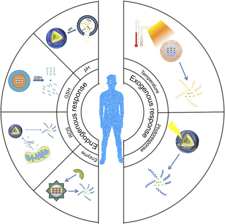
Tumor microenvironment-responsive polypeptide nanogels belong to a biomaterial with excellent biocompatibility, easily adjustable performance, biodegradability, and non-toxic properties. They are developed for selective delivery of antitumor drugs into target organs to promote tumor cell uptake, which has become an effective measure of tumor treatment. Endogenous (such as reduction, reactive oxygen species, pH, and enzyme) and exogenous (such as light and temperature) responsive nanogels can release drugs in response to tumor tissues or cells to improve drug distribution and reduce drug side effects. This article systematically introduces the research progress in tumor microenvironment-responsive polypeptide nanogels to deliver antitumor drugs and provides a reference for the development of antitumor nanoformulations.

Keywords: copolymer; drug delivery; nanogels; nanoparticle; polypeptide; stimulus-responsive.


Conflict of interest statement

The authors declare that the research was conducted in the absence of any commercial or financial relationships that could be construed as a potential conflict of interest.

Figures

FIGURE 1
FIGURE 1
Schematic diagram of the environmentally responsive nanogels responding to environmental changes.
FIGURE 2
FIGURE 2
Physical properties of PLGA/CS and the action of drugs (Yan et al., 2017). (A) Synthesis method of nanogels. (B) Change in particle size of nanogels at different pH. (C) Endocytosis effect of nanogels. (D) Inhibition rate of MTX-loaded nanogel and MTX on tumor cells.
FIGURE 3
FIGURE 3
Formation of pH-responsive mPEG-b-PNLG nanogels and the mechanism underlying drug delivery (Li Y. et al., 2018).
FIGURE 4
FIGURE 4
Chemical structure of the PEG113-bP (Lys-DSA) polymer thiol reaction and in vitro and in vivo experimental characterizations (Yao et al., 2019). (A) Schematic diagram of the chemical structure of the polymer thiol reaction and the reduction-sensitive DOX release behavior after LMs/DOX endocytosis. (B) Hydrodynamic radius and TEM images of blank and drug-loaded nanogels. (C) Cytotoxicity of nanogels on L929 cells and MCF-7 cells in vitro. (D) Cytotoxicity of DOX-loaded nanogels and free DOX.
FIGURE 5
FIGURE 5
Schematic diagram of the nanogel system and cell experiments (Li S. et al., 2018). (A) Schematic diagram of the role of STP-NG/SHK in tumor and lung metastasis. (B) CLSM cell surface staining analysis of cell surface vimentin. (C) Semi-quantitative analysis of Figure B. (D) Immunologic evaluation of STP binding to cells with flow cytometry. (E) Cell uptake of NG/SHK-FITC and STP-NG/SHK-FITC. Western blot (F) and semi-quantitative analysis (G) of the cells.
FIGURE 6
FIGURE 6
Animal experiments regarding tumor inhibition by the nanogels (Li S. et al., 2018). (A) Tumor growth curve. (B) Hind limb tumors. (C) Average weight of the osteosarcoma. (D) Calculation of tumor necrosis size. (E) Hematoxylin–eosin staining of the primary tumor. (F) H&E staining of the lung metastases. (G) Appearance of the lungs in each group. (H) H&E staining of mouse organs.
FIGURE 7
FIGURE 7
Characterization of self-assembly process and drug release of the nanogels (Wang et al., 2012). (A) Self-assembly process and drug release of the nanogels. (B) TEM image and particle size distribution of nanogels and the scanning electron microscope image and particle size of nanogels in the tetrahydrofuran solution. (C) Drug release of PEG-PCys-PPhe. (D) Confocal laser scanning microscopy (CLSM) image regarding co-incubation of HeLa cells and PEG-PCys-PPhe.
FIGURE 8
FIGURE 8
Characterization and treatment results of in situ DCMs (Deepagan et al., 2016). (A) DCMs maintained stability in circulation and the schematic diagram regarding DCMs triggering drug release by lysis in the tumor tissue. (B) Size distribution of NCM and DCMs. (C) Tumor growth curve. (D) H &E staining images of tumor tissue.
FIGURE 9
FIGURE 9
Synthesis and biological characterization of nanogels (Kim et al., 2013). (A) Schematic diagram of nanoparticle self-assembly. (B) Localization of DOX-loaded nanogels in cells. Effects of DOX-loaded nanogels on the tumor volume (C) and body weight (D).
FIGURE 10
FIGURE 10
Physical and cellular characterization of temperature-sensitive nanocarriers (Ko et al., 2015). (A) Schematic diagram of temperature-sensitive nanocarriers. (B) ζ potential of nanogels prepared by PEG-PK-PA and HA). (C) Relationship between the particle size distribution of nanogels and different temperatures (blue curve for 20°Cand brown curve for 37°C). (D) Endocytosis after incubation for 12 h. (E) Relationship between the cytotoxicity of PEG-PK-PA/HA and the concentration of nanogels. (F) After inhibitor treatment, fluorescence-activated cell sorting data were compared to analyze the internalization of fluorescein isothiocyanate–conjugated bovine serum albumin–loaded nanogels with zero ζ potential (ζ0). Zero potential of ζ+, ζ0 , and ζ was +47 mV, 0, and −47 mV, respectively.
FIGURE 11
FIGURE 11
Schematic diagram of the nanogel structure. Such a structure can enhance cellular uptake and nuclear delivery of drugs, thereby contributing to the effective circumvention of multidrug resistance in cancer (Chen et al., 2019).

Similar articles

Cited by

References

    1. Abdullah-Al-Nahain N., Nam J. A., Mok H., Lee Y.-k., Park S. Y. (2013). Dual-responsive Crosslinked Pluronic Micelles as a Carrier to Deliver Anticancer Drug Taxol. Macromol. Res. 21 (1), 92–99. 10.1007/s13233-013-1011-z - DOI
    1. Arroyo-Crespo J. J., Armiñán A., Charbonnier D., Balzano-Nogueira L., Huertas-López F., Martí C., et al. (2018). Tumor Microenvironment-Targeted Poly-L-Glutamic Acid-Based Combination Conjugate for Enhanced Triple Negative Breast Cancer Treatment. Biomaterials 186, 8–21. 10.1016/j.biomaterials.2018.09.023 - DOI - PubMed
    1. Bae Y., Kataoka K. (2009). Intelligent Polymeric Micelles from Functional Poly(ethylene Glycol)-Poly(amino Acid) Block Copolymers. Adv. Drug Deliv. Rev. 61 (10), 768–784. 10.1016/j.addr.2009.04.016 - DOI - PubMed
    1. Bahadur K C R., Xu P. (2012). Multicompartment Intracellular Self-Expanding Nanogel for Targeted Delivery of Drug Cocktail. Adv. Mater. 24 (48), 6479–6483. 10.1002/adma.201202687 - DOI - PubMed
    1. Bawa K. K., Oh J. K. (2017). Stimulus-Responsive Degradable Polylactide-Based Block Copolymer Nanoassemblies for Controlled/Enhanced Drug Delivery. Mol. Pharm. 14 (8), 2460–2474. 10.1021/acs.molpharmaceut.7b00284 - DOI - PubMed

LinkOut - more resources