. 2021 Oct 27:12:751667.
doi: 10.3389/fphar.2021.751667. eCollection 2021.

Angiogenesis is Inhibited by Arsenic Trioxide Through Downregulation of the CircHIPK3/miR-149-5p/FOXO1/VEGF Functional Module in Rheumatoid Arthritis

Affiliations

Angiogenesis is Inhibited by Arsenic Trioxide Through Downregulation of the CircHIPK3/miR-149-5p/FOXO1/VEGF Functional Module in Rheumatoid Arthritis

Juan Zhang et al. Front Pharmacol. .

Abstract

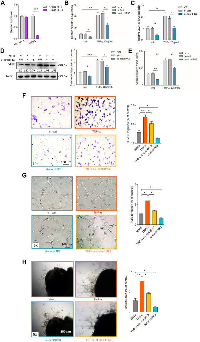
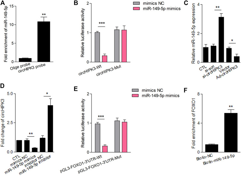
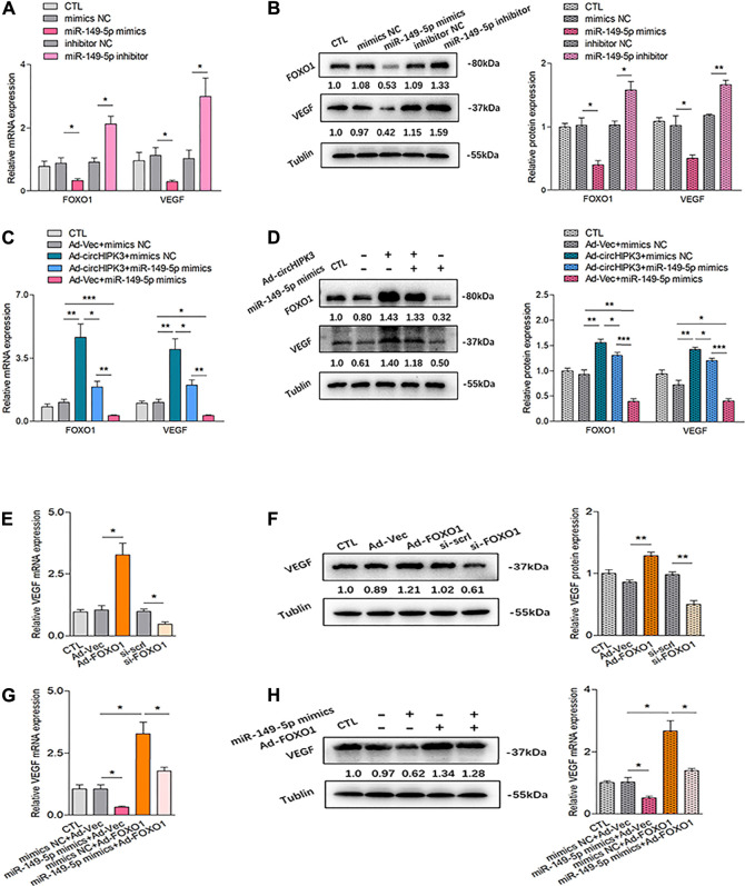
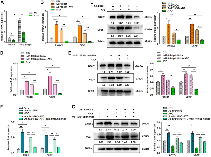
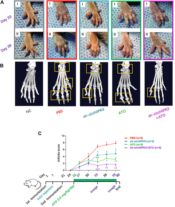
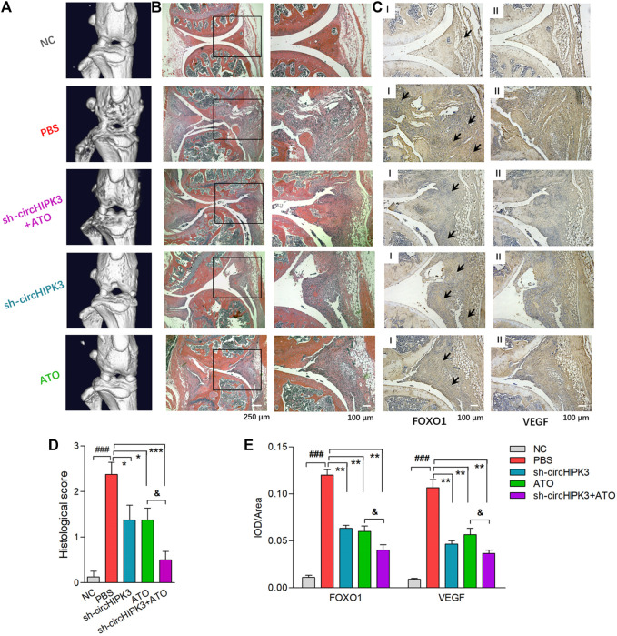
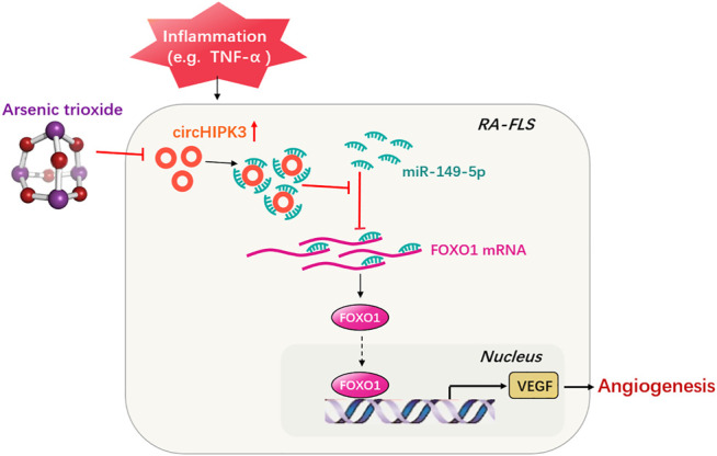
Angiogenesis is a crucial event in the pathogenesis of rheumatoid arthritis (RA). Arsenic trioxide (ATO, As2O3) has been reported to inhibit synovial angiogenesis via the vascular endothelial growth factor (VEGF)-centered functional module. However, the exact mechanisms of ATO on VEGF modulation remain unclear. Circular RNAs (circRNAs) are emerging as important regulators in RA, and the detailed mechanisms remain largely unknown. Here, we reported a circRNA (circHIPK3), the expression of which was significantly increased in RA fibroblast-like synoviocytes (RA-FLS) after TNF-α induction. Moreover, VEGF content in the supernatants of a RA-FLS and human dermal microvascular endothelial cell (HDMEC) co-culture as well as in RA-FLS co-cultured was significantly elevated in accordance with circHIPK3 levels. This increased VEGF expression may significantly upregulate endothelial tube formation and transwell migration, as well as microvessel sprouting in the ex vivo aortic ring assay. CircHIPK3 was further illustrated to be a sponge for the forkhead box transcription factor O1 (FOXO1)-targeting miR-149-5p, leading to the changing expression of the downstream VEGF. These networked factors mainly form a functional module regulating angiogenesis in RA-FLS, and the expression of this functional module could be significantly downregulated by ATO with a consistently reduced vascularity in vitro. In the collagen-induced arthritis (CIA) mice model, an intra-articular injection of the adeno-associated virus-si-circHIPK3 or ATO was demonstrated to alleviate the synovial VEGF expression and arthritis severity respectively. Thus, we elucidate a previously unknown mechanism between circRNAs and RA, and ATO has a significant protective effect on RA-FLS and CIA synovium via its inhibition of the angiogenic functional module of circHIPK3/miR-149-5p/FOXO1/VEGF, suggesting great potential for the combination therapy of ATO with circHIPK3 silencing.

Keywords: FoxO1; VEGF; angiogenesis; circHIPK3; inflammation; rheumatoid arthritis.


Conflict of interest statement

The authors declare that the research was conducted in the absence of any commercial or financial relationships that could be construed as a potential conflict of interest.

Figures

FIGURE 1
FIGURE 1
Overexpression of circHIPK3 in RA-FLS induced by TNF-α, and knockdown of circHIPK3 inhibited the VEGF production of RA-FLS. (A) CircHIPK3 could resist the digestion of Rnase R (n = 3; ***p < 0.001). (B) The expression of circHIPK3 could be significantly upregulated by TNF-α induction for 48 h, and significantly reduced by circHIPK3 knockdown whether TNF-α addition was performed or not (n = 3; *p < 0.05, **p < 0.01). (C–E) The effects of circHIPK3 silencing on VEGF mRNA (C) and protein (D) expression in co-cultured RA-FLS; VEGF concentration in co-culture’s supernatants (E) were determined by qRT-PCR, Western blot, and ELISA, respectively. VEGF expression was significantly upregulated after TNF-α induction, and significantly downregulated by circHIPK3 knockdown compared to those from the control group whether TNF-α was added or not (n = 3; *p < 0.05, **p < 0.01, ***p < 0.001). (F–H) The transwell assay (F) and tube formation test (G) for 6 h as well as ex vivo aortic ring angiogenesis assay (H) demonstrated significant changes in migration, the capillary-like structure formation of HDMECs and microvessel sprouting, respectively, in accordance with VEGF expression in supernatants from RA-FLS and HDMEC co-cultures (n = 3; *p < 0.05, **p < 0.01). Results are expressed as the mean ± S.E.M. Veh = vehicle control. CTL = PBS control group. si-scrl = scramble siRNA. TNF-α = 50 ng/ml.
FIGURE 2
FIGURE 2
CircHIPK3 interacted with miR-149-5p, and FOXO1 was a target of miR-149-5p. (A) MiR-149-5p was pulled down and enriched with biotinylated circHIPK3 probe (n = 3; **p < 0.01). (B) The relative luciferase activity of circHIPK3 was markedly suppressed when co-transfected with wild circHIPK3 and miR-149-5p mimics compared with the control reporter or mutated luciferase reporter (n = 3; ***p < 0.001). (C) qRT–PCR detection showed that the expression of miR-149-5p was significantly increased/reduced in the circHIPK3 knockdown/overexpression group compared with each control group (n = 3; *p < 0.05, **p < 0.01). (D) The level of circHIPK3 could be significantly reduced/enhanced by miR-149-5p mimics/inhibitor compared with each control group (n = 3; *p < 0.05, **p < 0.01). (E) Luciferase reporter assays showed that the luciferase activity of FOXO1 wild-type reporter was significantly reduced when transfected with miR-149-5p mimics compared with control reporter or mutated luciferase reporter (n = 3; ***p < 0.001). (F) A significant enrichment of FOXO1 mRNA in the pulled down sediments of 3′ end biotin-labeled miR-149-5p. (n = 3; **p < 0.01). Results are expressed as the mean ± S.E.M. CTL = PBS control group. si-scrl = scramble siRNA. NC = negative control.
FIGURE 3
FIGURE 3
Confirmation of the circHIPK3/miR-149-5p/FOXO1/VEGF functional module in vitro. (A–B) FOXO1/VEGF mRNA (A) and protein expression (B) were obviously down-/up-regulated by miR-149-5p mimics/inhibitor (n = 3; *p < 0.05, **p < 0.01). (C–D) Co-effects of circHIPK3 and miR-149-5p on FOXO1/VEGF expression in RA-FLS were evaluated. FOXO1/VEGF mRNA (C) and protein. (D) The expressions were significantly elevated/reduced by circHIPK3 overexpression/miR-149-5p mimics compared with those of each control group. Moreover, the significant downregulation of FOXO1/VEGF expression repressed by miR-149-5p mimics could be obviously rescued by circHIPK3 overexpression; in other words, the significant upregulation of FOXO1/VEGF expression induced by circHIPK3 overexpression could be significantly inhibited by miR-149-5p mimics (n = 3; *p < 0.05, **p < 0.01, ***p < 0.001). (E–F) The mRNA (E) and protein (F) expression of VEGF in RA-FLS were significantly elevated/reduced by FOXO1 overexpression/knockdown (n = 3; *p < 0.05, **p < 0.01). (G–H) FOXO1 overexpression significantly upregulated VEGF mRNA (G) and protein (H) expression, and the increment of VEGF expression induced by FOXO1 overexpression could be significantly suppressed by miR-149-5p mimics (n = 3; *p < 0.05). Results are expressed as the mean ± S.E.M. CTL = PBS control group. si-scrl = scramble siRNA. NC = negative control.
FIGURE 4
FIGURE 4
ATO inhibited angiogenesis via modulating the circHIPK3/miR-149-5p/FOXO1/VEGF functional module in RA-FLS in vitro. (A) After RA-FLS were exposed to 50 ng/ml TNF-α and 2.0 µM ATO for 48h, the expression of circHIPK3 was upregulated by TNF-α induction, and this upregulation could be significantly suppressed by ATO treatment (n = 3; *p < 0.05). (B–C) The expression of FOXO1/VEGF mRNA (B) and protein (C) were significantly upregulated by FOXO1 overexpression and downregulated by ATO. Furthermore, the FOXO1/VEGF downregulation induced by ATO could be rescued partially by FOXO1 overexpression (n = 3; *p < 0.05, **p < 0.01). (D–E) The expression of FOXO1/VEGF mRNA (D) and protein (E) were significantly upregulated by miR-149-5p inhibitor, and downregulated by ATO. Additionally, the FOXO1/VEGF expression downregulated by ATO could be partially rescued by miR-149-5p inhibitor (n = 3; *p < 0.05, **p < 0.01, ***p < 0.001). (F–G) The expression of FOXO1/VEGF mRNA (F) and protein (G) were significantly upregulated by circHIPK3 overexpression, and downregulated by ATO. Moreover, the expression of FOXO1/VEGF downregulated by ATO could be partially rescued by circHIPK3 overexpression but significantly reduced again further upon miR-149-5p mimics addition (n = 3; *p < 0.05, **p < 0.01, ***p < 0.001). Results are expressed as the mean ± S.E.M. CTL = PBS control group.
FIGURE 5
FIGURE 5
CircHIPK3 knockdown or its combined application with ATO alleviated the symptoms and joint destruction of CIA mice. (A) Representative pictures of the paws of mice showed that CIA developed rapidly in paws after the mice were immunized with collagen II and sustained to the end of the experiment. AAV-sh-circHIPK3, 2.0 mg/kg/day ATO administration, or the combination treatment of above two agents significantly alleviated the severity of arthritis compared with the CIA group on day 33 (Ⅰ) and day 39 (Ⅱ) after the first immunization. (B) The micro-CT images of paws demonstrated the destruction of the bone and cartilage in CIA mice, and ATO injection alleviated the joint destruction. AAV-sh-circHIPK3 intra-articular therapy in addition to ATO could evidently reduce the damages of bone compared with ATO therapy alone. (C) Clinical evaluation and the process diagram are demonstrated. Clinical arthritis scores showed that both ATO and intra-articular AAV-sh-circHIPK3 therapy could significantly reduce the arthritis scores of CIA mice. Moreover, combining ATO and AAV-sh-circHIPK3 reduced the arthritis score more significantly than ATO treatment alone (n = 5; *p < 0.05, **p < 0.01, ***p < 0.001 versus PBS group). Data are expressed as the mean ± SEM.
FIGURE 6
FIGURE 6
Histological evaluation of single or combined treatment of AAV-sh-circ-HIPK3 and ATO. (A) Micro-CT images of knee joints demonstrated the destruction of bone and cartilage in CIA mice, and ATO injection alleviated the joint destruction. AAV-sh-circHIPK3 combined with ATO could significantly reduce the damages of bone compared to ATO therapy alone. (B) The severity of arthritis in CIA mice was assessed by H&E staining of knee joint sections. ATO treatment at 2.0 mg/kg/day and AAV-sh-circHIPK3 injection, respectively, significantly reduced synovial hyperplasia, cartilage/bone erosion and joint inflammation compared with CIA control mice. Additionally, the combination therapy of ATO and AAV-sh-circHIPK3 yielded more reduction of pannus formation and inflammatory infiltration. (C) Immunohistochemical analysis was performed to evaluate the effect of AAV-sh-circHIPK3 and ATO on FOXO1 (Ⅰ) and VEGF (Ⅱ) expression in synovial tissues of CIA mice. CIA mice under PBS treatment had significantly increased staining intensity of FOXO1 and VEGF compared with normal mice. And the injection of AAV-sh-circHIPK3 or ATO significantly decreased staining for FOXO1 and VEGF compared with CIA control mice. Interestingly, FOXO1 and VEGF staining in the combined treatment of AAV-sh-circ-HIPK3 and ATO group were significantly weaker than those in the CIA mice under ATO therapy alone. Histological scores (D) and average IOD values for FOXO1 and VEGF immunostaining in the knee joint synovium (E) were, respectively, evaluated and shown (n = 5; *p < 0.05, **p < 0.01, ***p < 0.001 versus PBS treatment group; ###p < 0.001 versus NC; and p < 0.05 versus ATO group). Data are expressed as the mean ± SEM. NC = normal control group.
FIGURE 7
FIGURE 7
Schematic illustration underlying the mechanism of ATO’s effect on the circHIPK3/miR-149-5p/FOXO1/VEGF functional module. CircHIPK3 might act as a sponge for the FOXO1-targeting miR-149-5p, leading to the downregulation of downstream VEGF. These networked factors mainly form a functional module regulating angiogenesis in RA-FLS, and the expression of this functional module could be significantly downregulated by ATO, ultimately inhibiting angiogenesis.

References

    1. Altesha M. A., Ni T., Khan A., Liu K., Zheng X. (2019). Circular RNA in Cardiovascular Disease. J. Cel Physiol. 234 (5), 5588–5600. 10.1002/jcp.27384 - DOI - PubMed
    1. Capitão M., Soares R. (2016). Angiogenesis and Inflammation Crosstalk in Diabetic Retinopathy. J. Cel Biochem. 117 (11), 2443–2453. 10.1002/jcb.25575 - DOI - PubMed
    1. Chen L. L. (2020). The Expanding Regulatory Mechanisms and Cellular Functions of Circular RNAs. Nat. Rev. Mol. Cel Biol. 21 (8), 475–490. 10.1038/s41580-020-0243-y - DOI - PubMed
    1. Chu M., Zhang C. (2018). Inhibition of Angiogenesis by Leflunomide via Targeting the Soluble Ephrin-A1/EphA2 System in Bladder Cancer. Sci. Rep. 8 (1), 1539. 10.1038/s41598-018-19788-y - DOI - PMC - PubMed
    1. Dai X., Chen C., Yang Q., Xue J., Chen X., Sun B., et al. (2018). Exosomal circRNA_100284 From Arsenite-Transformed Cells, via microRNA-217 Regulation of EZH2, Is Involved in the Malignant Transformation of Human Hepatic Cells by Accelerating the Cell Cycle and Promoting Cell Proliferation. Cell Death Dis. 9 (5), 454. 10.1038/s41419-018-0485-1 - DOI - PMC - PubMed