. 2021 Oct 28:12:686530.
doi: 10.3389/fimmu.2021.686530. eCollection 2021.

Highly Purified Alloantigen-Specific Tregs From Healthy and Chronic Kidney Disease Patients Can Be Long-Term Expanded, Maintaining a Suppressive Phenotype and Function in the Presence of Inflammatory Cytokines

Affiliations

Highly Purified Alloantigen-Specific Tregs From Healthy and Chronic Kidney Disease Patients Can Be Long-Term Expanded, Maintaining a Suppressive Phenotype and Function in the Presence of Inflammatory Cytokines

Arimelek Cortés-Hernández et al. Front Immunol. .

Abstract

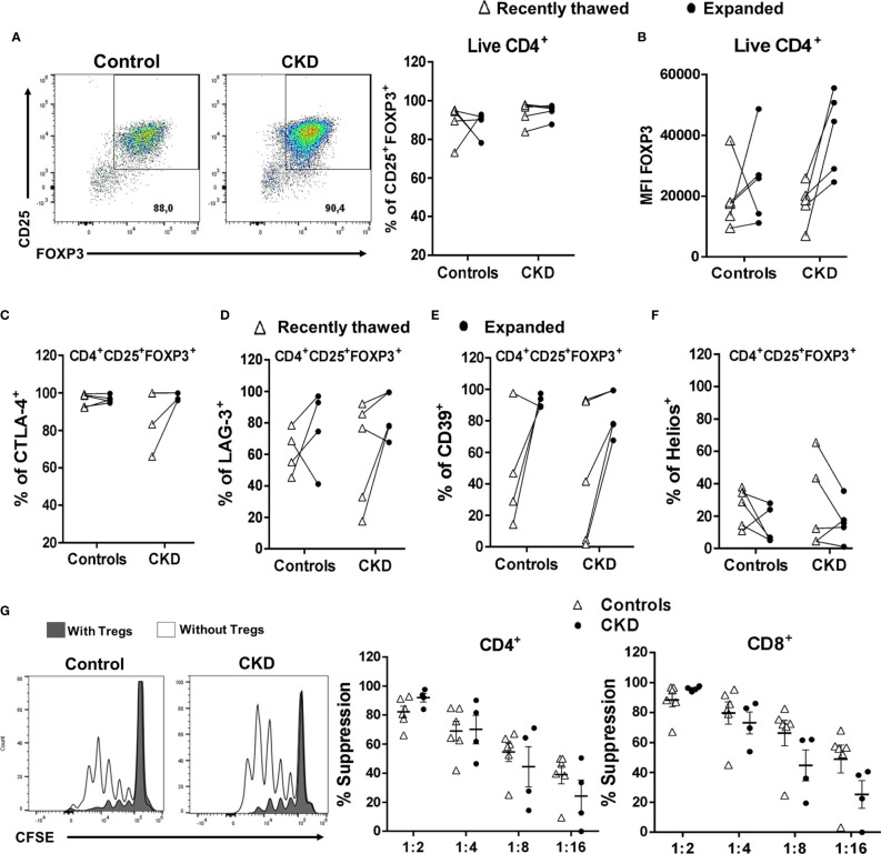
The adoptive transfer of alloantigen-specific regulatory T cells (alloTregs) has been proposed as a therapeutic alternative in kidney transplant recipients to the use of lifelong immunosuppressive drugs that cause serious side effects. However, the clinical application of alloTregs has been limited due to their low frequency in peripheral blood and the scarce development of efficient protocols to ensure their purity, expansion, and stability. Here, we describe a new experimental protocol that allows the long-term expansion of highly purified allospecific natural Tregs (nTregs) from both healthy controls and chronic kidney disease (CKD) patients, which maintain their phenotype and suppressive function under inflammatory conditions. Firstly, we co-cultured CellTrace Violet (CTV)-labeled Tregs from CKD patients or healthy individuals with allogeneic monocyte-derived dendritic cells in the presence of interleukin 2 (IL-2) and retinoic acid. Then, proliferating CD4+CD25hiCTV- Tregs (allospecific) were sorted by fluorescence-activated cell sorting (FACS) and polyclonally expanded with anti-CD3/CD28-coated beads in the presence of transforming growth factor beta (TGF-β), IL-2, and rapamycin. After 4 weeks, alloTregs were expanded up to 2,300 times the initial numbers with a purity of >95% (CD4+CD25hiFOXP3+). The resulting allospecific Tregs showed high expressions of CTLA-4, LAG-3, and CD39, indicative of a highly suppressive phenotype. Accordingly, expanded alloTregs efficiently suppressed T-cell proliferation in an antigen-specific manner, even in the presence of inflammatory cytokines (IFN-γ, IL-4, IL-6, or TNF-α). Unexpectedly, the long-term expansion resulted in an increased methylation of the specific demethylated region of Foxp3. Interestingly, alloTregs from both normal individuals and CKD patients maintained their immunosuppressive phenotype and function after being expanded for two additional weeks under an inflammatory microenvironment. Finally, phenotypic and functional evaluation of cryopreserved alloTregs demonstrated the feasibility of long-term storage and supports the potential use of this cellular product for personalized Treg therapy in transplanted patients.

Keywords: allospecific; expansion; regulatory T cells; suppression; transplantation.


Conflict of interest statement

The authors declare that the research was conducted in the absence of any commercial or financial relationships that could be construed as a potential conflict of interest.

Figures

Figure 1
Figure 1
Schematic representation of the protocol used for ex vivo expansion of alloantigen-specific regulatory T cells (Tregs). CD4+CD25hiCD127 T cells [labeled with CellTrace Violet (CTV)] sorted using fluorescence-activated cell sorting (FACS) were stimulated with allogeneic monocyte-derived dendritic cells (Mo-DCs) in the presence of IL-2 and retinoic acid. On day 7, CD4+CD25hiCTV T cells (allospecific Tregs) were purified by FACS, rested for 3 days, and then polyclonally expanded for 4 days stimulated with anti-CD3/anti-CD28, IL-2, TGF-β, and rapamycin, followed by 3 days resting with only IL-2. After four rounds of polyclonal expansion, analysis of the phenotype and in vitro functional assays were performed.
Figure 2
Figure 2
Highly purified allospecific regulatory T cells (Tregs) increased the expressions of FOXP3 and CD25 after ex vivo expansion. CD4+CD25hiCD127 T cells [labeled with CellTrace Violet (CTV)] from healthy individuals (white triangles, n = 7) or patients with chronic kidney disease (CKD) (black circles, n = 5) were stimulated with allogeneic monocyte-derived dendritic cells (Mo-DCs). On day 7, CD4+CD25hiCTV T cells (allospecific Tregs) were purified and polyclonally expanded for 4 weeks; the expressions of FOXP3 and CD25 were evaluated by flow cytometry. (A) Allospecific Tregs from patients proliferated to the same extent as the Tregs from healthy controls. Fold expansion was calculated by dividing the number of Tregs obtained on the evaluated day by the number of Tregs on day 0. (B–D) The proportion of CD25+FOXP3+ cells (B) and the expressions of FOXP3 (C) and CD25 (D) were increased in the expanded alloantigen-specific Tregs (alloTregs) in both study groups compared to those of freshly isolated Tregs. The median fluorescence intensity (MFI) was calculated as described in Section 2.6. Representative experiments are shown in (BD), and the white histograms represent FMO (fluorescence minus one) controls (C, D). The results are shown as the mean ± SEM. Statistical analysis was performed using the Mann–Whitney U test or Wilcoxon’s rank-sum test.
Figure 3
Figure 3
Long-term expanded alloantigen-specific regulatory T cells (alloTregs) had high expressions of CTLA-4, LAG-3, and CD39. Highly purified alloTregs from healthy controls (white triangles, n = 5-7) or patients with chronic kidney disease (CKD) (black circles, n = 5) were polyclonally expanded for 4 weeks and then were stained with fluorochrome-conjugated antibodies for analysis by flow cytometry. (A–D) Analysis of typical Treg markers showed that the expanded alloTregs from both healthy individuals and patients with CKD had high proportions of CTLA-4+ (A), LAG-3+ (B), CD39+ (C), and Helios+ (D) cells. Representative experiments are shown (A–D), and the white histograms represent FMO (fluorescence minus one) controls. The results are shown as the mean ± SEM. Statistical analysis was performed using the Mann–Whitney U test. No significant differences were observed.
Figure 4
Figure 4
Expanded allospecific regulatory T cells (Tregs) suppressed the proliferation of conventional T cells in an alloantigen-specific manner. Expanded allospecific Tregs (alloTregs) from healthy individuals (white triangles, n = 7) or patients with chronic kidney disease (CKD) (black circles, n = 5) were co-cultured with conventional CD3+ T cells [labeled with carboxy fluorescein succinimidyl ester (CFSE)] and stimulated with allogeneic monocyte-derived dendritic cells (Mo-DCs) from their respective donors or from non-related individuals (third party). On day 4 of culture, T-cell proliferation was evaluated by flow cytometry. (A) Representative experiment. (B) Expanded alloTregs suppressed the proliferation of both CD8+ and CD4+ conventional T cells (Tconv) only when they were stimulated with the dendritic cells (DCs) with which they were initially expanded (DCs from donor), but not when they were stimulated with unrelated DCs (third party). (C) alloTregs efficiently suppressed alloreactive T cells in the presence of the inflammatory cytokines IFN-γ, IL-4, IL-6, and TNF-α (C). The relative increase (IR) of the percentage of suppression was calculated by dividing the value in the presence of the cytokine by the value in the absence of cytokine (medium). All experiments were performed in duplicate. The results are shown as the mean ± SEM. Statistical analysis was performed using unpaired t-test (B) or one-way ANOVA (B).
Figure 5
Figure 5
Long-term expansion reduced the demethylation of TSDR-Foxp3 in purified allospecific regulatory T cells (Tregs). CpG methylation analysis of the Treg-specific demethylated region (TSDR) in the Foxp3 gene was evaluated in purified CD4+CD25hiCD127 Tregs (freshly isolated) and in expanded alloantigen-specific Tregs (alloTregs). (A) Each square represents one CpG site analyzed (of a total of 15 CpGs). Six to seven clones per DNA sample were sequenced. The demethylation color code ranges from white (0% demethylation) to navy blue (80% demethylation), according to the color scale. Purified CD4+CD25CD45RA+ T cells (naive) displayed a completely methylated TSDR and were used as the negative control. (B) Freshly isolated Tregs from both groups displayed high demethylation of TSDR-Foxp3, while expanded alloTregs showed a significant increase in the methylation of the TSDR. DNA samples from three male chronic kidney disease (CKD) patients and four male healthy controls were analyzed. The results are expressed as the mean ± SEM. Statistical analysis was performed using paired t-test.
Figure 6
Figure 6
Expanded alloantigen-specific regulatory T cells (Tregs) maintain their immunosuppressive phenotype after stimulation in an inflammatory microenvironment. Long-term expanded Tregs from healthy controls (white triangles, n = 7) or patients with chronic kidney disease (CKD) (black circles, n = 5) were stimulated with anti-CD3/anti-CD28 in the presence or absence of inflammatory cytokines (IFN-γ, IL-6, IL-4, or TNF-α) for two additional weeks. The expressions of Treg markers were determined by flow cytometry. (A–D) Activation of allospecific Tregs from CKD patients in the presence inflammatory cytokines did not have an effect on the percentages of CD25+FOXP3+ (A, B), CTLA-4+ (D), and Helios+ (E) cells and on the expression of FOXP3 (C) to the same extent as the Tregs from healthy controls. A representative experiment is shown in (A). The relative increase (IR) of the percentage or median fluorescence intensity (MFI) was calculated by dividing the value in the presence of the cytokine by the value in the absence of cytokines (medium). The results are shown as the mean ± SEM. Statistical analysis was performed using the Kruskal–Wallis test. No significant differences were observed.
Figure 7
Figure 7
Allospecific regulatory T cells (alloTregs) maintained their in vitro function after stimulation in an inflammatory microenvironment. Long-term expanded alloTregs from healthy individuals (white triangles, n = 5) or patients with chronic kidney disease (CKD) (black circles, n = 5) were stimulated with anti-CD3/anti-CD28 in the presence or absence of inflammatory cytokines for two additional weeks, and then in vitro allospecific suppression assays were performed. (A, B) The alloTregs from both patients and healthy controls maintained their capacity to suppress both CD4+ (left) and CD8+ (right) T-cell proliferation after expansion in the presence of all the inflammatory cytokines evaluated (IFN-γ, IL-6, IL-4, and TNF-α). A representative experiment is shown in (A). The relative increase (IR) of the percentage of suppression was calculated by dividing the value in the presence of the cytokine by the value in the absence of cytokines (medium). All experiments were performed in duplicate. The results are shown as the mean ± SEM. Statistical analysis was performed using the Kruskal–Wallis test. No significant differences were observed.
Figure 8
Figure 8
Expanded regulatory T cells (Tregs) maintained their phenotype and in vitro function after long-term cryopreservation. On day 28 of polyclonal expansion, alloantigen-specific Tregs (alloTregs) from healthy individuals (white triangles, n = 4) or patients with chronic kidney disease (CKD) (black circles, n = 4) were cryopreserved for a long time (>6 months), and then the cells were thawed and polyclonally expanded. The expressions of the Tregs markers and their in vitro immunosuppressive function were determined by flow cytometry. (A–F) After 2 weeks of stimulation, thawed alloTregs from both groups maintained the proportions of CD25+FOXP3+ (A), CTLA-4+ (C), LAG-3+ (D), and CD39+ (E) cells and the expression of FOXP3 (B), but a low proportion of Helios+ cells (F). (G) Cryopreserved allospecific Tregs can suppress the proliferation of both polyclonal CD4+ (left) and CD8+ (right) T cells after 2 weeks of expansion. Representative experiments are shown in (A, G). The results are shown as the mean ± SEM. Statistical analysis was performed using the Mann–Whitney U test or Wilcoxon test. No significant differences were observed.
Figure 9
Figure 9
Cryopreserved allospecific regulatory T cells (alloTregs) expressed high levels of the chemokine receptors CXCR3, CCR4, and CCR2. Long-term cryopreserved alloTregs from healthy controls (white triangles, n = 4) or patients with chronic kidney disease (CKD) (black circles, n = 4) were thawed and polyclonally expanded for 2 weeks. The expressions of chemokine receptors were determined by flow cytometry. (A, B) The expanded Tregs of both groups had high percentages of CXCR3+ (A), CCR4+ (B), and CCR2+ (C) cells, but a low proportion of CCR7+ cells (D). Representative experiments are shown in (A–D). White histograms represent FMO (fluorescence minus one) controls. The results are shown as the mean ± SEM. Statistical analysis was performed using unpaired t-test. No significant differences were observed.

Similar articles

Cited by

References

    1. Vaidya SR, Aeddula NR. Chronic Renal Failure. Treasure Island (FL: StatPearls; (2019).
    1. Holt CD. Overview of Immunosuppressive Therapy in Solid Organ Transplantation. Anesthesiol Clinics (2017) 35(3):365–80. doi: 10.1016/j.anclin.2017.04.001 - DOI - PubMed
    1. Cohen-Bucay A, Gordon CE, Francis JM. Non-Immunological Complications Following Kidney Transplantation. F1000Res (2019) 8:F1000 Faculty Rev–194. doi: 10.12688/f1000research.16627.1 - DOI - PMC - PubMed
    1. Lu L, Barbi J, Pan F. The Regulation of Immune Tolerance by FOXP3. Nat Rev Immunol (2017) 17(11):703–17. doi: 10.1038/nri.2017.75 - DOI - PMC - PubMed
    1. Hu M, Wang C, Zhang GY, Saito M, Wang YM, Fernandez MA, et al. . Infiltrating Foxp3(+) Regulatory T Cells From Spontaneously Tolerant Kidney Allografts Demonstrate Donor-Specific Tolerance. Am J Transplant (2013) 13(11):2819–30. doi: 10.1111/ajt.12445 - DOI - PubMed

Publication types

MeSH terms