Review
. 2021 Sep 15;13(5):707-716.
doi: 10.1007/s12551-021-00831-8. eCollection 2021 Oct.

What keeps us ticking? Sinoatrial node mechano-sensitivity: the grandfather clock of cardiac rhythm

Affiliations
Review

What keeps us ticking? Sinoatrial node mechano-sensitivity: the grandfather clock of cardiac rhythm

Eilidh A MacDonald et al. Biophys Rev. .

Abstract

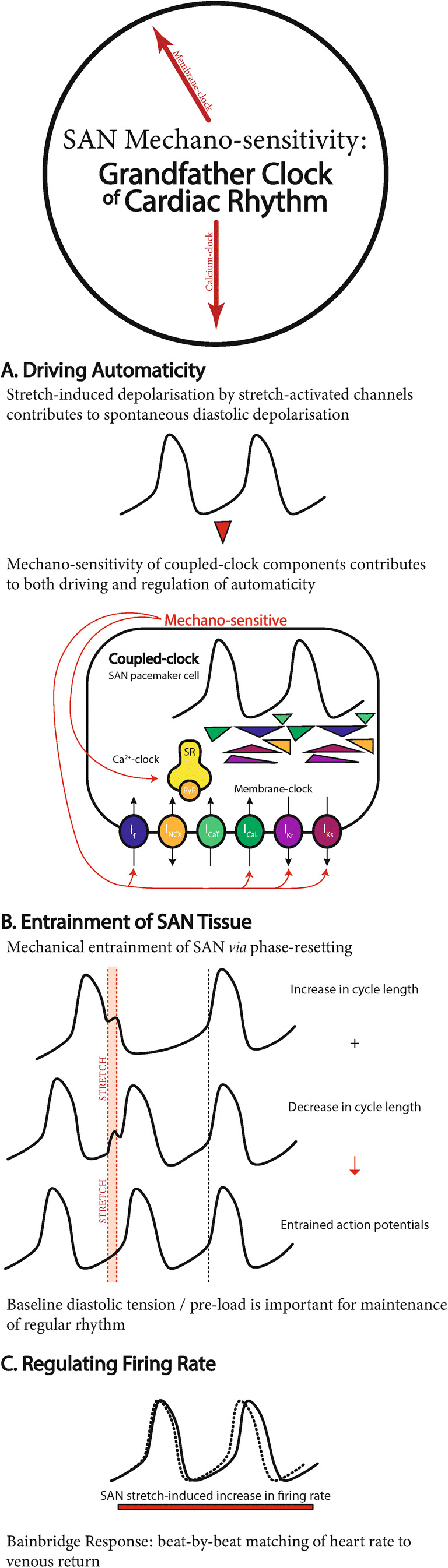
The rhythmic and spontaneously generated electrical excitation that triggers the heartbeat originates in the sinoatrial node (SAN). SAN automaticity has been thoroughly investigated, which has uncovered fundamental mechanisms involved in cardiac pacemaking that are generally categorised into two interacting and overlapping systems: the 'membrane' and 'Ca2+ clock'. The principal focus of research has been on these two systems of oscillators, which have been studied primarily in single cells and isolated tissue, experimental preparations that do not consider mechanical factors present in the whole heart. SAN mechano-sensitivity has long been known to be a contributor to SAN pacemaking-both as a driver and regulator of automaticity-but its essential nature has been underappreciated. In this review, following a description of the traditional 'clocks' of SAN automaticity, we describe mechanisms of SAN mechano-sensitivity and its vital role for SAN function, making the argument that the 'mechanics oscillator' is, in fact, the 'grandfather clock' of cardiac rhythm.

Keywords: Calcium clock; Heart rate; Mechano-electric coupling; Membrane clock; Pacemaking; Stretch.


Figures

Fig. 1
Fig. 1
The grandfather clock of cardiac rhythm. Summary of the role of mechano-sensitivity in sinoatrial node (SAN) A automaticity, B entrainment, and C regulation. For expanded figures of the coupled-clock system, please refer to Lakatta et al. (2010), Quinn and Kohl (2012), and Bartos et al. (2015)
Fig. 2
Fig. 2
Stretch-induced increase in the beating rate of isolated sinoatrial node preparations. A Intracellular sharp electrode recording of transmembrane potential (top) and applied and generated force (bottom; passive stretch and active contraction pointing upwards) in spontaneously beating cat isolated sinoatrial node (SAN) tissue (from Deck 1964) and B patch-clamp recording of transmembrane potential in a spontaneously beating rabbit isolated SAN cell (light curve, before stretch; dark curve, during stretch) (from Cooper et al. 2000). Both show an increase in beating rate during stretch, accompanied by a reduction in the absolute value of maximum diastolic and maximum systolic potential
Fig. 3
Fig. 3
Phase resetting in the sinoatrial node (SAN). A Application of subthreshold square-wave pulse in the early (1), middle (2), and late (3) phase of the cardiac cycle in the rabbit isolated SAN (lower tracings in each section are action potentials from the SAN region close to the atrium to show time of stimulus artefacts) and B the relationship between cycle length and time of stimulation in the cardiac cycle, showing that subthreshold depolarising current pulses result in an increase or a decrease in cycle length, depending on the timing of the stimulation within the cardiac cycle. From Sano et al. (1978)
Fig. 4
Fig. 4
Effects of physiological levels of baseline stretch on isolated sinoatrial node (SAN) beating rate. Floating microelectrode recordings of transmembrane potential in cat isolated SAN, showing a stretch-induced shift of the maximum diastolic potential towards less negative values, resulting in A restoration of regular rhythm in a SAN with irregular activity at slack length or B initiation of spontaneous excitation in a previously quiescent SAN. In both examples, tissue length was increased by ~40% from slack, with periods of stretch indicated by the lower horizontal lines. From Lange et al. (1966)

Similar articles

Cited by

References

    1. Abramovich-Sivan S, Akselrod S (1999) Phase response curve based model of the SA node: simulation by two-dimensional array of pacemaker cells with randomly distributed cycle lengths. Med Biol Eng Comput 37:482–491. 10.1007/BF02513334 - PubMed
    1. Anumonwo JMB, Delmar M, Vinet A, Michfaels DC, Jalife J (1991) Phase resetting and entrainment of pacemaker activity in single sinus nodal cells. Circ Res 68:1138–1153. 10.1161/01.res.68.4.1138 - PubMed
    1. Arai A, Kodama I, Toyama J (1996) Roles of Cl- channels and Ca2+ mobilization in stretch-induced increase of SA node pacemaker activity. Am J Physiol Heart Circ Physiol 270:H1726–H1735. 10.1152/ajpheart.1996.270.5.H1726 - PubMed
    1. Bainbridge FA (1915) The influence of venous filling upon the rate of the heart. J Physiol 50:65–84. 10.1113/jphysiol.1915.sp001736 - PMC - PubMed
    1. Bartos DC, Grandi E, Ripplinger CM (2015) Ion channels in the heart. Compr Physiol 5:1423–1464. 10.1002/cphy.c140069 - PMC - PubMed