. 2021 Sep 22;12(40):13425-13433.
doi: 10.1039/d1sc04464h. eCollection 2021 Oct 20.

Targeted protein oxidation using a chromophore-modified rapamycin analog

Affiliations

Targeted protein oxidation using a chromophore-modified rapamycin analog

Taylor M Courtney et al. Chem Sci. .

Abstract

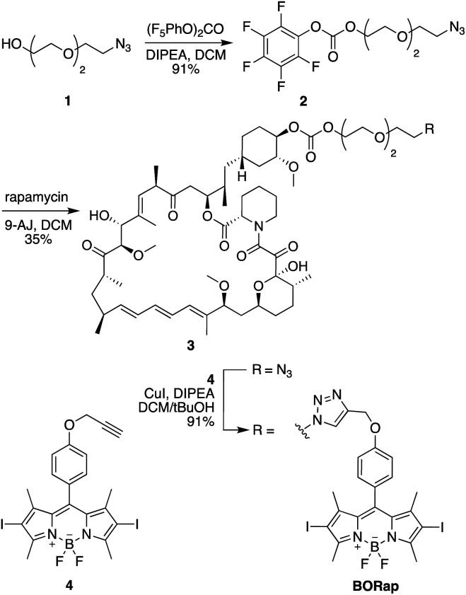
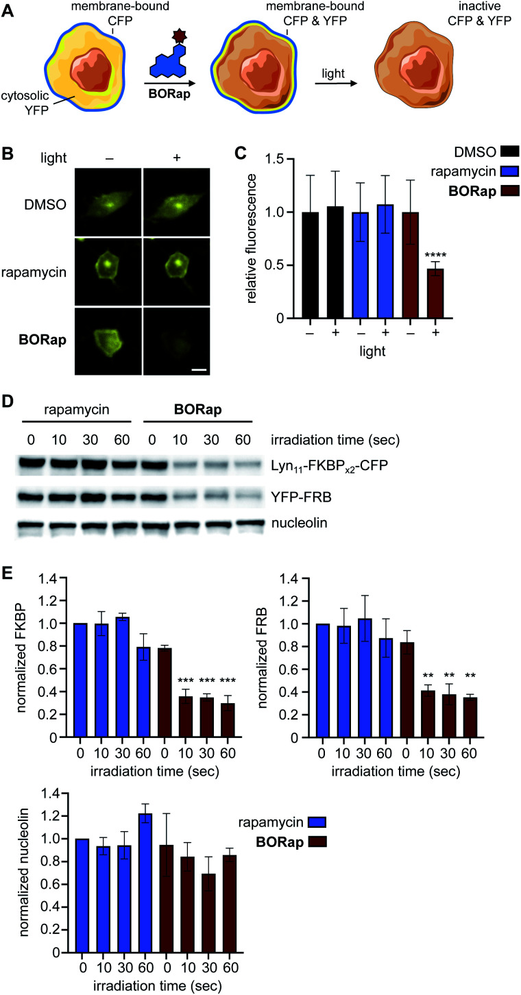
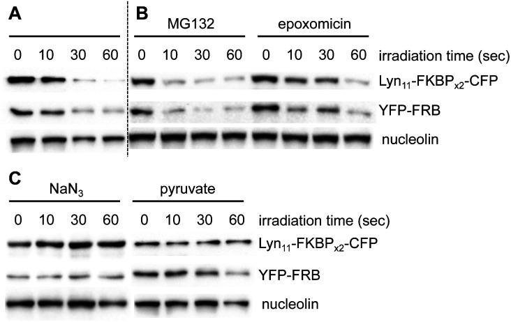
Chemically induced dimerization of FKBP and FRB using rapamycin and rapamycin analogs has been utilized in a variety of biological applications. Formation of the FKBP-rapamycin-FRB ternary complex is typically used to activate a biological process and this interaction has proven to be essentially irreversible. In many cases, it would be beneficial to also have temporal control over deactivating a biological process once it has been initiated. Thus, we developed the first reactive oxygen species-generating rapamycin analog toward this goal. The BODIPY-rapamycin analog BORap is capable of dimerizing FKBP and FRB to form a ternary complex, and upon irradiation with 530 nm light, generates singlet oxygen to oxidize and inactivate proteins of interest fused to FKBP/FRB.


Conflict of interest statement

There are no conflicts of interest to declare.

Figures

Fig. 1
Fig. 1. Synthesis of the BODIPY-modified rapamycin analog BORap.
Fig. 2
Fig. 2. (A) Schematic representation of chemically induced protein dimerization and activation of luciferase, followed by subsequent light exposure and resulting deactivation of the enzyme. (B) We found that 100 nM of rapamycin and 10 μM of BORap elicited optimal activity, and as such, were used in the subsequent experiment. (C) A decrease in luminescence was observed with increasing irradiation times for BORap-treated cells, while no light-induced effect was seen in the rapamycin control. Average signal intensity is plotted with error bars representing standard deviation. Statistical analysis of triplicate experiments was performed in Prism using a two-way ANOVA test to compare irradiated samples for each treatment condition to non-irradiated (0 s) samples. P values less than or equal to 0.05 are represented by *, less than or equal to 0.0001 are represented by ****. Comparisons of rapamycin-treated samples (10, 30, 60, or 90 s vs. 0 s) and comparison of BORap-treated samples (10 s vs. 0 s) are not significant.
Fig. 3
Fig. 3. (A) Schematic representation of the split-TEV assay. (B) HEK293T cells expressing pFKBP-TEVpCT, pFRB-TEVpNT, and PCS2-GFP-TEV were treated with either rapamycin or BORap and irradiated to monitor change in fluorescence intensity. Cells were irradiated through a DsRed filter for 0 or 90 seconds, and post-irradiation images were acquired after 18 hours. Scale bar equals 50 μM. (C) Average fluorescence intensity of 20 cells for each condition was quantified and normalized to the DMSO negative control, with error bars representing standard deviations. Statistical analysis was performed in Prism using a one-way ANOVA test to compare each treatment condition to the DMSO negative control. P values less than or equal to 0.0001 are represented by ****, while the irradiated BORap sample shows no statistical difference from the DMSO negative control.
Fig. 4
Fig. 4. (A) Schematic representation of the membrane reporter translocation assay. (B) Cells treated with either rapamycin or BORap and irradiated at 0 hours followed by YFP imaging at 2 hours. Scale bar equals 10 μM. (C) Quantification of the YFP signal intensity before and after irradiation (90 s), showed reduced fluorescence in BORap-treated cells, while all other conditions show little to no decrease in fluorescent signal. Average fluorescence intensity in 20 cells is shown normalized to pre-irradiated controls, with error bars representing standard deviations. Statistical analysis was performed in Prism using a two-way ANOVA test to compare irradiated samples for each treatment condition to pre-irradiated samples. P values less than or equal to 0.0001 are represented by ****, while all other comparisons are not significant. (D) HEK293T cells expressing the CFP and YFP constructs were treated with 20 μM of rapamycin or BORap prior to irradiating with 530 nm light. Western blot analysis using anti-GFP, anti-GAPDH, and anti-nucleolin antibodies showed a light-induced band decrease with BORap but not rapamycin treatment. (E) Triplicate experiments were performed and quantified using FIJI. Band intensity of FKBP, FRB, and nucleolin for each irradiation timepoint were normalized to the rapamycin-treated, non-irradiated sample and averaged. Statistical analysis was performed as above, comparing irradiated to non-irradiated samples within each treatment condition (10, 30, or 60 s vs. 0 s). P values less than or equal to 0.001 are represented by ***, less than or equal to 0.01 by **, while comparisons of rapamycin-treated samples (for FKBP, FRB, and nucleolin) and comparison of BORap-treated samples (for nucleolin) are not significant.
Fig. 5
Fig. 5. (A) A western blot was performed as above with BORap-treated cells used as a control to show inactivation of the membrane reporter. (B) Treatment with MG132 or epoxomicin shows no change, suggesting that reduced band intensity is not the result of proteasomal protein degradation. (C) Sodium azide functions to block loss of signal, while sodium pyruvate has less or no effect, thus supporting that BORap-induced signal loss requires the generation of singlet oxygen.

Similar articles

References

    1. Wang P. Gao Z. Yuan M. Zhu J. Wang F. Mechanically linked poly[2]rotaxanes constructed from the benzo-21-crown-7/secondary ammonium salt recognition motif. Polym. Chem. 2016;7(22):3664–3668. doi: 10.1039/C6PY00494F. - DOI
    1. Voß S. Klewer L. Wu Y. W. Chemically induced dimerization: reversible and spatiotemporal control of protein function in cells. Curr. Opin. Chem. Biol. 2015;28:194–201. doi: 10.1016/j.cbpa.2015.09.003. - DOI - PubMed
    1. Wu H. D. Kikuchi M. Dagliyan O. Aragaki A. K. Nakamura H. Dokholyan N. V. Umehara T. Inoue T. Rational design and implementation of a chemically inducible heterotrimerization system. Nat. Methods. 2020;17(9):928–936. doi: 10.1038/s41592-020-0913-x. - DOI - PMC - PubMed
    1. Williams D. J. Puhl III H. L. Ikeda S. R. Rapid Modification of Proteins Using a Rapamycin-Inducible Tobacco Etch Virus Protease System. PLoS One. 2009;4(10):e7474. doi: 10.1371/journal.pone.0007474. - DOI - PMC - PubMed
    1. Diaz J. E. Morgan C. W. Minogue C. E. Hebert A. S. Coon J. J. Wells J. A. A Split-Abl Kinase for Direct Activation in Cells. Cell Chem. Biol. 2017;24(10):1250–1258. doi: 10.1016/j.chembiol.2017.08.007. - DOI - PMC - PubMed