. 2021 Sep 16;12(40):13477-13482.
doi: 10.1039/d1sc03065e. eCollection 2021 Oct 20.

Real-time imaging of cell-surface proteins with antibody-based fluorogenic probes

Affiliations

Real-time imaging of cell-surface proteins with antibody-based fluorogenic probes

Wenchao Wang et al. Chem Sci. .

Abstract

Cell-surface proteins, working as key agents in various diseases, are the targets for around 66% of approved human drugs. A general strategy to selectively detect these proteins in a real-time manner is expected to facilitate the development of new drugs and medical diagnoses. Although brilliant successes were attained using small-molecule probes, they could cover a narrow range of targets due to the lack of suitable ligands and some of them suffer from selectivity issues. We report herein an antibody-based fluorogenic probe prepared via a two-step chemical modification under physiological conditions, to fulfill the selective recognition and wash-free imaging of membrane proteins, establishing a modular strategy with broad implications for biochemical research and for therapeutics.


Conflict of interest statement

There are no conflicts to declare.

Figures

Fig. 1
Fig. 1. Strategy of antibody-based fluorogenic probes (Ab-PnSRB, n indicates the PEG length). (a) Schematic illustration of cell-surface protein imaging with antibody-based probes prepared by two-step labeling (details are shown in Scheme S3†). (b) The representative conformations of 2PCA–PEGn/SRB-N3 (PnSRB in short) labeled necitumumab (Nec) at the stable trajectories. Average distances and SDs between the centroid of the xanthene of two SRBs were calculated. (c) Labeling efficiency of 2PCA–PEGn/SRB-N3 towards antibodies (i.e., necitumumab and human IgG), which was visualized by in-gel fluorescent scanning (FL).
Fig. 2
Fig. 2. Photophysical properties of SRB-N3 and the resulting Ab-PnSRB probes. (a) Fluorescence spectra of SRB-N3 (5 μM) in different solvents (λex = 530 nm). (b) Fluorescence spectra of Nec-PnSRB (100 nM) and SRB-N3 (20 nM) in H2O (λex = 530 nm). (c) Fluorescence response of Nec-P5SRB/IgG-P5SRB (20 nM) in the presence of recombinant EGFR at different ratios (λex = 560 nm and λem = 590 nm).
Fig. 3
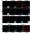
Fig. 3. Live-cell imaging by using Ab-PnSRB (λex = 561 nm and λem = 581–639 nm). (a) Cellular performance of Nec-PnSRB (100 nM) in both EGFR-positive (A431) and EGFR-negative (HepG2) cells. 10× Nec means that cells were pretreated with 1 μM of necitumumab for 0.5 h before the addition of Nec-P5SRB. Control probes IgG-PnSRB (100 nM) were studied in parallel. (b) Comparison of the background signal from IgG-P5SRB and that from IgG-P5TER (“always-on” probe) in A431 cells without washing.
Fig. 4
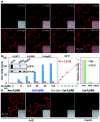
Fig. 4. Applications of Ab-P5SRB for detecting endogenous EGFR in living cells. (a) Real-time imaging of A431 cells upon addition of Nec-P5SRB (100 nM). (b) FACS of Nec-P5SRB towards A431/A549/HepG2 (left). Western blotting (WB) analysis of EGFR in A431/A549/HepG2 (inset). Linear correlation analysis between the average fluorescence in A431 and probe's concentration (middle). Relative intensity readings extracted from WB (pink) versus those from FACS (green) (right). (c) No-wash imaging of A431/HepG2 cells obtained by using Cet-P5SRB (100 nM). 10 × Cet in the third panel means that cells were pretreated with 1 μM of cetuximab for 0.5 h before the addition of Cet-P5SRB. λex = 561 nm and λem = 581–639 nm for all cell imaging.

References

    1. Kuhlmann L. Cummins E. Samudio I. Kislinger T. Expert Rev. Proteomics. 2018;15:259–275. doi: 10.1080/14789450.2018.1429924. - DOI - PubMed
    1. Bausch-Fluck D. Goldmann U. Müller S. van Oostrum M. Müller M. Schubert O. T. Wollscheid B. Proc. Natl. Acad. Sci. U. S. A. 2018;115:E10988–E10997. doi: 10.1073/pnas.1808790115. - DOI - PMC - PubMed
    1. Leung K. K. Wilson G. M. Kirkemo L. L. Riley N. M. Coon J. J. Wells J. A. Proc. Natl. Acad. Sci. U. S. A. 2020;117:7764–7775. doi: 10.1073/pnas.1917947117. - DOI - PMC - PubMed
    1. Keppler A. Gendreizig S. Gronemeyer T. Pick H. Vogel H. Johnsson K. Nat. Biotechnol. 2003;21:86–89. doi: 10.1038/nbt765. - DOI - PubMed
    1. Gao J. Hori Y. Nishiura M. Bordy M. Hasserodt J. Kikuchi K. Chem. Lett. 2020;49:232–235. doi: 10.1246/cl.190875. - DOI