. 2021 Sep 6;12(40):13492-13505.
doi: 10.1039/d1sc02775a. eCollection 2021 Oct 20.

Modelling the active SARS-CoV-2 helicase complex as a basis for structure-based inhibitor design

Affiliations

Modelling the active SARS-CoV-2 helicase complex as a basis for structure-based inhibitor design

Dénes Berta et al. Chem Sci. .

Abstract

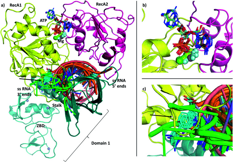
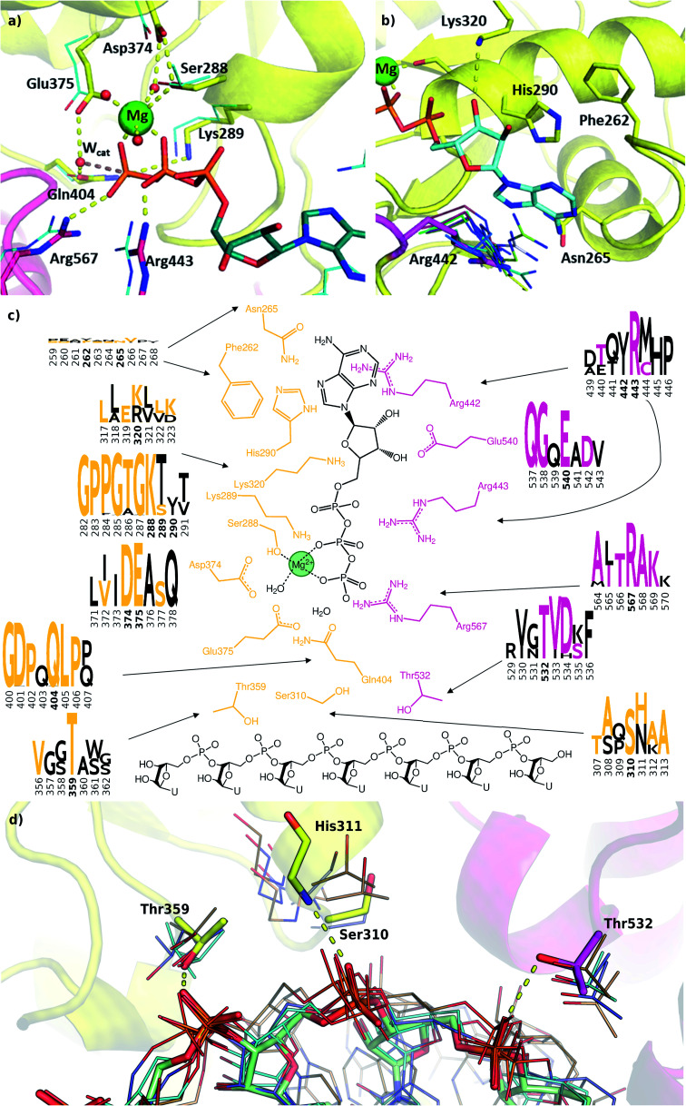
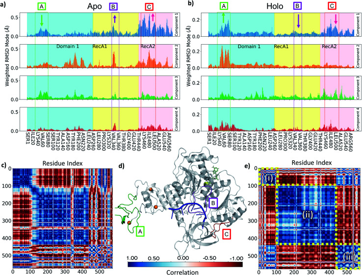
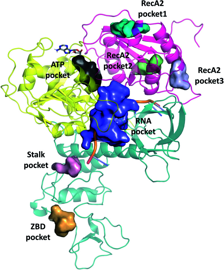
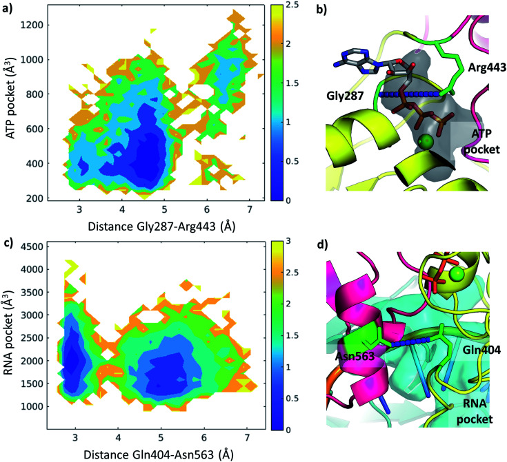
The RNA helicase (non-structural protein 13, NSP13) of SARS-CoV-2 is essential for viral replication, and it is highly conserved among the coronaviridae family, thus a prominent drug target to treat COVID-19. We present here structural models and dynamics of the helicase in complex with its native substrates based on thorough analysis of homologous sequences and existing experimental structures. We performed and analysed microseconds of molecular dynamics (MD) simulations, and our model provides valuable insights to the binding of the ATP and ssRNA at the atomic level. We identify the principal motions characterising the enzyme and highlight the effect of the natural substrates on this dynamics. Furthermore, allosteric binding sites are suggested by our pocket analysis. Our obtained structural and dynamical insights are important for subsequent studies of the catalytic function and for the development of specific inhibitors at our characterised binding pockets for this promising COVID-19 drug target.


Conflict of interest statement

There are no conflicts to declare.

Figures

Fig. 1
Fig. 1. (a) Our model of the RNA helicase NSP13 of SARS-CoV-2 monomer (cartoon) coloured by three domains: RecA1 (yellow), RecA2 (magenta), and Domain 1 (aquamarine). ATP analogues (sticks) along with Mg (green sphere) and single stranded nucleic acids are depicted from aligned homologous structures (full list of PDB codes are available in Table S1†). 3′ ends of the nucleic acids present the same orientation in all chains (highlighted in green). (b) Position of the ATP analogues (nucleotides in stick and metal ions and compounds in spheres) in homologous structures. (c) Specific helicase inhibitor binding region with allosteric inhibitors displayed in cyan (black arrow).
Fig. 2
Fig. 2. Structural comparison of the deposited PDB structures of the helicase dimer in SARS-CoV-1 (PDBID: 6jyt, a), SARS-CoV-2 (PDBID: 6zsl, b), SARS-CoV-2 in complex with NSP7 NSP8 and NSP12 (PDBID: 6xez, c) and SARS-CoV-2 with a small fragment of ssRNA bound in complex with NSP7 NSP8 and NSP12 (PDBID: 7cxm, d). The interaction between the two helicase monomers differs depending on the experimental method used to resolve the structures.
Fig. 3
Fig. 3. (a) Distribution of the identity of sequences in multiple sequence alignment compared to the SARS-CoV-2 helicase. There are only members of coronaviridae above 300 matching residues (50%, lime circles, 52 entries). There are no sequences with medium similarity (107–300 similar residues, red circles). The mass of the sequences matches only 107 residues or less (blue circles). The closest relatives (96 sequences represented by 52) are grouped in coronavirus subfamilies (grouped in the y axis) with principal hosts highlighted in the inset. (b) Sequence identity of the representative sequences of 796 clusters from UniProtKB aligned to the 601-residue long SARS-CoV-2 RNA helicase. Domain 1 shows similarity only to the close relatives (52 sequences, representing clusters of 96), while the RecA1 and RecA2 domains are more common across ATPase sequences. Key structural motifs are highlighted using symbols (P-loop: grey square, DE motif: green square, arginine fingers: black triangle, ssRNA interactions: red triangles).
Fig. 4
Fig. 4. Modelling and conservation of the ATP and RNA sites. (a) Main protein-substrate interactions of the triphosphate and magnesium ions are compared with alignment for PDB template 2xzo (cyan lines). (b) Nucleotide-binding region focusing on Arg442 (magenta sticks) is aligned with homologous arginine residues (lines, PDB structures 5k8u, 5vhc, 5xdr, 5y4z, 5 y6m, 5y6n, 6adx, 6ady, 6c90 and 6jim). (c) Sequence conservation for RecA1 (orange) and RecA2 (magenta) domains are depicted in logos for each residue and its neighbours (data from Fig. 3b). Coloured letters represent the residues in the SARS-CoV-2 helicase sequence, depicted residue indices are bold in the logos. (d) Structures of the RNA binding region aligned with existing RNA-helicase crystal structures complexed with ssRNA (depicted in lines). RecA1 and RecA2 domains are shown in yellow and magenta, respectively. Key residues (sticks) are labelled, and H-bonds are depicted in yellow dashes.
Fig. 5
Fig. 5. (a) 2D free energy profile along with the puckering angle and the distance between uracil 6 (OP1) and threonine 359 (OG), from all Amber holo simulations. The colour bar represents the hight of the free energy profile in kcal mol−1. Insets depict the structures in the three local minima, showing uracil 6 in grey sticks and threonine 359 as yellow sticks. Specific distance between the residues is highlighted by yellow dashes. (b and c) Distribution of the puckering angle along the MD simulations using CHARMM (blue) and Amber (orange) force field for uracil 3 (b) and 6 (c).
Fig. 6
Fig. 6. (a and b) Comparison of PCA Weighted-RMSD scores holo monomer (a) and apo monomer (b) simulations with Amber. (c) DCC map of holo monomer simulation with Amber. (d) Key areas highlighted in the helicase structure (d, grey cartoon) and on panels a and b (text box and arrows indicating relative change in magnitude): (A) Zinc Binding region (green), (B) Domain 1 loop from Ile334 to Gln354 (purple), (C) RecA1/2 interface loop from Thr450 to Ala510 (red). Bound objects are shown and coloured: RNA in blue (centre), ATP in lime (upper right), zinc in orange (left spheres) and magnesium as a yellow sphere. (e) DCC map of apo monomer simulation with Amber for motions described by the first principal component. Colour bar providing correlation scale is shown in the bottom centre.
Fig. 7
Fig. 7. Overview of binding pockets (coloured surfaces) identified in the holo complex MD trajectories. ATP (grey sticks) and the ssRNA (cartoon) are only show for the sake of orientation.
Fig. 8
Fig. 8. Free energy profiles depicted along selected coordinates and substrate pocket volumes (holo Amber trajectories). (a) 2D profile along Gly287-Arg443 distance and ATP pocket volume. The color bar represents the height of the free energy profile in kcal mol−1. (b) The ATP pocket (black surface) and the Gly287-Arg443 distance (residues in green, distance in blue) depicted in a representative structure. (c) 2D profile along Gln404-Asn563 distance and RNA pocket volume. The color bar represents the height of the free energy profile in kcal mol−1. (d) The RNA pocket (cyan surface) and the Gln404-Asn563 distance (residues in green, distance in blue) depicted in a representative structure.

Similar articles

Cited by

References

    1. Wang M. Yan M. Xu H. Liang W. Kan B. Zheng B. Chen H. Zheng H. Xu Y. Zhang E. Wang H. Ye J. Li G. Li M. Cui Z. Liu Y.-F. Guo R.-T. Liu X.-N. Zhan L.-H. Zhou D.-H. Zhao A. Hai R. Yu D. Guan Y. Xu J. Emerging Infect. Dis. 2005;11:1860–1865. doi: 10.3201/eid1112.041293. - DOI - PMC - PubMed
    1. Wu F. Zhao S. Yu B. Chen Y.-M. Wang W. Song Z.-G. Hu Y. Tao Z.-W. Tian J.-H. Pei Y.-Y. Yuan M.-L. Zhang Y.-L. Dai F.-H. Liu Y. Wang Q.-M. Zheng J.-J. Xu L. Holmes E. C. Zhang Y.-Z. Nature. 2020;579:265–269. doi: 10.1038/s41586-020-2008-3. - DOI - PMC - PubMed
    1. Beigel J. H. Tomashek K. M. Dodd L. E. Mehta A. K. Zingman B. S. Kalil A. C. Hohmann E. Chu H. Y. Luetkemeyer A. Kline S. Lopez de Castilla D. Finberg R. W. Dierberg K. Tapson V. Hsieh L. Patterson T. F. Paredes R. Sweeney D. A. Short W. R. Touloumi G. Lye D. C. Ohmagari N. Oh M. Ruiz-Palacios G. M. Benfield T. Fätkenheuer G. Kortepeter M. G. Atmar R. L. Creech C. B. Lundgren J. Babiker A. G. Pett S. Neaton J. D. Burgess T. H. Bonnett T. Green M. Makowski M. Osinusi A. Nayak S. Lane H. C. N. Engl. J. Med. 2020;383:1813–1826. doi: 10.1056/NEJMoa2007764. - DOI - PMC - PubMed
    1. Chen J. Malone B. Llewellyn E. Grasso M. Shelton P. M. M. Olinares P. D. B. Maruthi K. Eng E. Vatandaslar H. Chait B. T. Kapoor T. Darst S. A. Campbell E. A. Cell. 2020;182:1560–1573. doi: 10.1016/j.cell.2020.07.033. - DOI - PMC - PubMed
    1. Wang Y. Zhang D. Du G. Du R. Zhao J. Jin Y. Fu S. Gao L. Cheng Z. Lu Q. Hu Y. Luo G. Wang K. Lu Y. Li H. Wang S. Ruan S. Yang C. Mei C. Wang Y. Ding D. Wu F. Tang X. Ye X. Ye Y. Liu B. Yang J. Yin W. Wang A. Fan G. Zhou F. Liu Z. Gu X. Xu J. Shang L. Zhang Y. Cao L. Guo T. Wan Y. Qin H. Jiang Y. Jaki T. Hayden F. G. Horby P. W. Cao B. Wang C. Lancet. 2020;395:1569–1578. doi: 10.1016/S0140-6736(20)31022-9. - DOI - PMC - PubMed