. 2021 Nov 5;11(4):20458940211056806.
doi: 10.1177/20458940211056806. eCollection 2021 Oct-Dec.

Evidence supporting a role for circulating macrophages in the regression of vascular remodeling following sub-chronic exposure to hemoglobin plus hypoxia

Affiliations

Evidence supporting a role for circulating macrophages in the regression of vascular remodeling following sub-chronic exposure to hemoglobin plus hypoxia

Vijaya Karoor et al. Pulm Circ. .

Erratum in

Abstract

Macrophages are a heterogeneous population with both pro- and anti-inflammatory functions play an essential role in maintaining tissue homeostasis, promoting inflammation under pathological conditions, and tissue repair after injury. In pulmonary hypertension, the M1 phenotype is more pro-inflammatory compared to the M2 phenotype, which is involved in tissue repair. The role of macrophages in the initiation and progression of pulmonary hypertension is well studied. However, their role in the regression of established pulmonary hypertension is not well known. Rats chronically exposed to hemoglobin (Hb) plus hypoxia (HX) share similarities to humans with pulmonary hypertension associated with hemolytic disease, including the presence of a unique macrophage phenotype surrounding distal vessels that are associated with vascular remodeling. These lung macrophages are characterized by high iron content, HO-1, ET-1, and IL-6, and are recruited from the circulation. Depletion of macrophages in this model prevents the development of pulmonary hypertension and vascular remodeling. In this study, we specifically investigate the regression of pulmonary hypertension over a four-week duration after rats were removed from Hb + HX exposure with and without gadolinium chloride administration. Withdrawal of Hb + HX reversed systolic pressures and right ventricular function after Hb + Hx exposure in four weeks. Our data show that depleting circulating monocytes/macrophages during reversal prevents complete recovery of right ventricular systolic pressure and vascular remodeling in this rat model of pulmonary hypertension at four weeks post exposure. The data presented offer a novel insight into the role of macrophages in the processes of pulmonary hypertension regression in a rodent model of Hb + Hx-driven disease.

Keywords: heart; hemoglobinopathies; lung; pulmonary vascular disease; sickle cell disease.


Figures

Fig. 1.
Fig. 1.
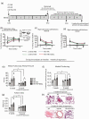
Cardiopulmonary analysis of the progression and the macrophage contribution in the resolution of hemoglobin (Hb) plus hypoxia (HX) mediated pulmonary hypertension. (a) Schematic of study design. Five weeks of exposure to Hb plus Hx followed by a four-week observational period after removal of Hb plus HX. (b–d) Echocardiography measurements of the pulmonary artery acceleration time and right ventricular wall thickness in diastole and systole at five weeks after progressive PH, and at two and four weeks after removal PH stimulus. (e–g) End point measurements of mean pulmonary artery pressure, pulmonary artery medial thickening, and right ventricular hypertrophy, and in a subset of rats sacrificed at five weeks and rats sacrificed after four weeks of recovery. (h) Microscopy visualization of H&E stained lung tissue in rats after five weeks of progressive PH (HX and Hb plus HX exposure) and after four weeks of recover. Normoxic rats were sacrificed at five weeks (n = 3) and nine weeks (n = 3). p < 0.05 - five-week time point vs. seven- and nine-week time point; ††p < 0.05 Hb + HX and Hb + HX + GdCl3 vs. HX and NX cohorts at nine-week time point; #p < 0.05 vs. NX at five weeks; *p < 0.05- five-week vs. nine-week time points; ǂp < 0.05 Hb + HX vs. Hb + HX+GdCl3 at nine-week time point; ¶ p < 0.05 NX vs. all other groups at nine-week time point.
Fig. 2.
Fig. 2.
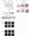
Lung iron accumulation and presence of cd68 and cd163 cell populations after five weeks of progressive PH and following four weeks of recovery. (a) Perls lung iron accumulation original magnification 60× Brown- iron, Black arrows- iron loaded cells (macrophages); (b) Western blot analysis of whole lung cd68; (c) mRNA quantification of cd163 in whole lung tissue; (d) Immunohistochemistry microscopy visualization for cells expressing cd163 in lung tissue sections of rats. Original magnification 40×. Red- cells expressing cd163. White arrows- positive cells.
Fig. 3.
Fig. 3.
Quantification and location of lung IL-6 and ET-1: (a) Western blot quantification of IL-6; (b) representative western blot of IL-6; (c) Immunohistochemistry visualization of location of lung and IL-6. Original magnification 40×. (d) Western blot quantification of ET-1; (e) representative western blot of ET-1; (f) Immunohistochemistry visualization of location of lung and ET-1. Original magnification 20× Immunohistochemistry visualization of location of lung HO-1 and ET-1. Original magnification 40×. p < 0.05 vs. Hb + HX at four weeks of regression; *p < 0.05 vs. HB+HX at five weeks of progressive PH; **NX vs. Hb + HX by Students t test.
Fig. 4.
Fig. 4.
Quantification and location of lung heme oxygenase-1 (HO-1) and Masons Trichrome staining. (a) Western blot quantification of HO-1; (b) Representative western blot of HO-1; (c) Immunohistochemistry visualization of location of lung and HO-1. Original magnification 40×; (d) Masons trichrome staining; Blue- collagen. Original magnification 20×. p < 0.05 vs. Hb + HX at four weeks of regression; **NX vs. Hb + HX by Students t test.

Similar articles

Cited by

References

    1. Tuder RM, Stacher E, Robinson J, et al.. Pathology of pulmonary hypertension. Clin Chest Med 2013; 34: 639–650. - PubMed
    1. Simonneau G, Montani D, Celermajer DS, et al.. Haemodynamic definitions and updated clinical classification of pulmonary hypertension. Eur Respir J 2019; 53: 1801913. - PMC - PubMed
    1. Gordeuk VR, Castro OL, Machado RF. Pathophysiology and treatment of pulmonary hypertension in sickle cell disease. Blood 2016; 127: 820–828. - PMC - PubMed
    1. Rother RP, Bell L, Hillmen P, et al.. The clinical sequelae of intravascular hemolysis and extracellular plasma hemoglobin: a novel mechanism of human disease. JAMA 2005; 293: 1653–1662. - PubMed
    1. Buehler PW, Baek JH, Lisk C, et al. . Free hemolgobin induction of pulmonary vascular disease: evidence for and inflammatory mechanism. Am J Physiol Lung Cell Mol Physiol 2012; 303: 312–326. - PMC - PubMed

LinkOut - more resources