Review
. 2021 Oct 27:11:775349.
doi: 10.3389/fonc.2021.775349. eCollection 2021.

Syndecans and Their Synstatins: Targeting an Organizer of Receptor Tyrosine Kinase Signaling at the Cell-Matrix Interface

Affiliations
Review

Syndecans and Their Synstatins: Targeting an Organizer of Receptor Tyrosine Kinase Signaling at the Cell-Matrix Interface

Alan C Rapraeger. Front Oncol. .

Abstract

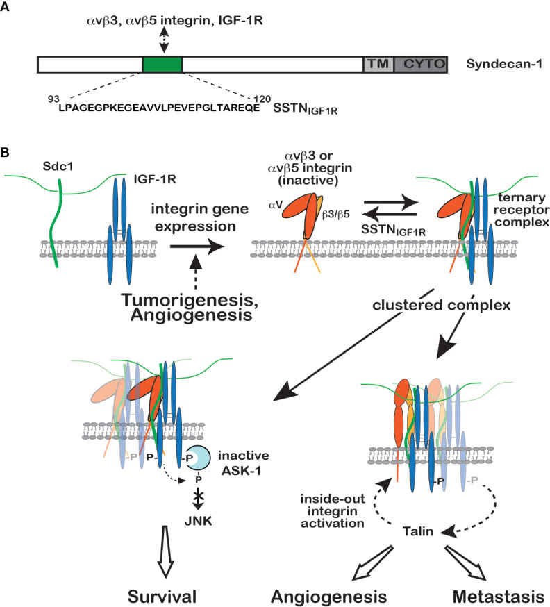
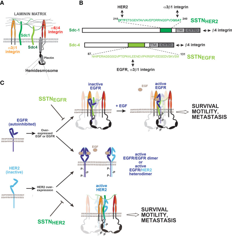
Receptor tyrosine kinases (RTKs) and integrin matrix receptors have well-established roles in tumor cell proliferation, invasion and survival, often functioning in a coordinated fashion at sites of cell-matrix adhesion. Central to this coordination are syndecans, another class of matrix receptor, that organize RTKs and integrins into functional units, relying on docking motifs in the syndecan extracellular domains to capture and localize RTKs (e.g., EGFR, IGF-1R, VEGFR2, HER2) and integrins (e.g., αvβ3, αvβ5, α4β1, α3β1, α6β4) to sites of adhesion. Peptide mimetics of the docking motifs in the syndecans, called "synstatins", prevent assembly of these receptor complexes, block their signaling activities and are highly effective against tumor cell invasion and survival and angiogenesis. This review describes our current understanding of these four syndecan-coupled mechanisms and their inhibitory synstatins (SSTNIGF1R, SSTNVEGFR2, SSTNVLA-4, SSTNEGFR and SSTNHER2).

Keywords: ASK-1; CXCR4; EGFR; IGF-1R; VEGFR2; integrin signaling; synstatin.


Conflict of interest statement

The author declares that the research was conducted in the absence of any commercial or financial relationships that could be construed as a potential conflict of interest.

Figures

Figure 1
Figure 1
Regulation of IGF-1R signaling by Sdc1. (A) Model depicting site of IGF-1R capture, along with either the αvβ3 or αvβ5 integrin, in the extracellular domain of human Sdc1. A peptide mimetic of this site, SSTNIGF1R, competitively blocks receptor binding. (B) IGF-1R is not engaged with Sdc1 in nontransformed cells, typically because they lack the αvβ3 and αvβ5 integrins (top left). But integrin expression, usually in response to malignant transformation or activation of endothelial cells during angiogenesis, results in integrin docking to the extracellular domain of Sdc1, which is followed by IGF-1R capture at the same docking site (top right). Once formed, constitutive or matrix-induced clustering of the ternary receptor complex activates IGF-1R by autophosphorylation that does not require IGF-1. Activated endothelial cells or tumor cells bearing the ternary receptor complex rely on the syndecan-activated IGF-1R to phosphorylate and suppress the activity of Apoptosis-Signal regulating Kinase-1 (ASK-1) engaged with the IGF-1R cytoplasmic domain (bottom left), preventing ASK-1-mediated activation of Jun-N-terminal Kinase (JNK) and blocking entry into apoptosis, thus promoting tumor cell survival. In a second activity (bottom right), downstream signaling from IGF-1R activates the αvβ3 or αvβ5 integrin via an inside-out signaling pathway that targets the integrin-activating protein Talin, resulting in endothelial or tumor cell motility during the onset of angiogenesis or tumor cell invasion. SSTNIGF1R competitively disrupts the ternary receptor complex, preventing integrin activation and removing the constitutive suppression of ASK-1. Neither activity can be rescued by IGF-1 when the receptor complex is disrupted by SSTNIGF1R, emphasizing the singular role played by the syndecan in this IGF-1R signaling mechanism (see discussion in text, references (, –41) and references therein.
Figure 2
Figure 2
Regulation of VLA-4 activation and polarized cell invasion by Sdc1. (A) VLA-4 and VEGFR2 docking sites in Sdc1. Juxtamembrane sites in the extracellular domain of human Sdc1 (DFTF and PVD) responsible for engaging very late antigen-4 (VLA-4, α4β1 integrin) and vascular endothelial growth factor receptor-2 (VEGFR2), respectively. Synstatin peptides containing one, but not both of these sites (e.g., SSTNVLA-4 or SSTNVEGFR2), prevent VLA-4 or VEGFR2 capture by the syndecan and disrupt signaling that relies on the co-capture of both receptors. (B) Regulation of high affinity VLA-4 adhesion by Sdc1. VLA-4 undergoes rapid activation when engaging ligand, involving a conformation change and clustering (avidity modulation) to increase binding affinity. On cells expressing Sdc1 (e.g., myeloma cells, vascular endothelial cells, melanoma, Jurkat-T cells), this activation is blocked by preventing VLA-4 docking with Sdc1 using SSTNVLA-4 or mutating the DFTF motif in the syndecan. The mechanism underlying this dependence on the syndecan remains under investigation. (C) Role of heparanase and shed Sdc1 in polarized cell invasion. The heparan-sulfate-degrading enzyme heparanase is a known tumor promoter and enhancer of leukocyte recruitment during inflammation. Trimming of the HS chains on Sdc1 exposes its core protein to cleavage by matrix-metalloproteinase-9 (MMP-9), releasing the syndecan ectodomain. The shed syndecan couples an inactive receptor complex consisting of VEGFR2 with inactive protein kinase A (PKA) attached to its cytoplasmic domain, the cytokine receptor CXCR4 and adenylate cyclase 7 (AC7) to the clustered integrin, causing VEGF-independent activation of VEGFR2; VEGFR2 phosphorylates CXCR4 on Y135, activating its Gαiβγ GTPase and the Gαi-dependent AC7. Local generation of cAMP leads to activation of protein kinase-A (PKA) engaged with the VEGFR2 cytoplasmic domain and phosphorylation of the α4-integrin cytoplasmic domain on serine 988. This displaces the Rac-inhibitory paxillin from the integrin, causing polarized invasion of VLA-4-dependent immune cells, typically tumor supporting cells such a macrophages, MDSCs and others, as well as myeloma cells. Displacement of paxillin also inhibits the cross-talk between VLA-4 and LFA-1 necessary for LFA-1-mediated migration that characterizes tumor suppressor cells, such as NK and cytotoxic T cells. Either prevention of integrin activation by SSTNVLA-4 or VEGFR2-coupling to the integrin by SSTNVEGFR2 serves to block these processes [see discussion in text, references (60, 64) and references therein].
Figure 3
Figure 3
Coupling of EGFR family members to integrins by Sdc1 and Sdc4 regulates wound healing, tumor cell invasion and survival. (A) Syndecans and integrins on quiescent epithelial or endothelial cells. The laminin-332 (LN332) binding α3β1 and α6β4 integrin, along with Sdc1 and Sdc4 via their heparan sulfate chains, mediate binding to LN332-rich basement membrane. The long (>1,000 amino acids) cytoplasmic domain of the β4 integrin subunit engages the plectrin and BP180 scaffolding proteins, which engage the intermediate filament network and help stabilize the incorporation of the α6β4 integrin into hemidesmosomes. (B) Sites of EGFR, HER2 and integrin capture in Sdc1 and Sdc4. The multifunctional juxtamembrane site in Sdc1 captures HER2 and the α3β1 integrin. Multiple binding interactions occur throughout the sequence; nonetheless, the DFTF motif that is also essential for binding VLA-4 plays a prominent role in HER2 binding, whereas α3β1 integrin capture is highly dependent on the QGAT motif. An analogous juxtamembrane site in Sdc4, which bears no homology to the site in Sdc1, captures EGFR and the α3β1 integrin, relying on multiple binding interactions throughout the sequence. Synstatin peptides based on these sites are highly selective for EGFR (SSTNEGFR) or HER2 (SSTNHER2) and displace α3β1 integrin only when coupled with the EGFR family member for which they are specific. In addition to these interactions, the C-termini of the syndecans engage the cytoplasmic C-terminus of the β4 integrin, which comprises the α6β4 integrin. This is also syndecan-type specific; mutations that disrupt Sdc1 binding having no effect on Sdc4 and vice versa. (C) Formation and activation of syndecan-organized quaternary receptor complexes containing EGFR or HER2. EGFR and HER2 are overexpressed in squamous cell carcinomas, along with EGF. EGFR depends on EGF for dimerization, and to relieve constraints in its extracellular domain that prevent dimerization. HER2 lacks these extracellular constraints but lacks a ligand to promote its dimerization. Relief of the inhibitory constraints in EGFR allows it to form active homodimers or active heterodimers with HER2. Activation of receptor tyrosine kinases causes the breakdown of hemidesmosomes, freeing the cytoplasmic domain of the β4 integrin to engage Sdc1 and Sdc4. Quaternary receptor complex is formed when EGFR and the α3β1 integrin, or HER2 and the α3β1 integrin assemble with their respective docking site on the syndecans as well. The kinases appear to be captured as monomers and rely on clustering of the complexes to sites of matrix adhesion to be activated. Simple clustering of the syndecan is sufficient to activate HER2, but EGFR also requires EGF, ostensibly to relieve the dimerization constraints in its extracellular domain. Several tyrosines in the β4 cytoplasmic domain become phosphorylated, and support signaling that leads to epithelial cell migration during wound healing, or invasion and survival signaling in tumor cells and endothelial cells engaged in pathological angiogenesis. Synstatin peptides specific for HER2 capture by Sdc1 (SSTNHER2) or EGFR capture by Sdc4 (SSTNEGFR) are highly specific inhibitors of these processes. [See discussion in text, references (–83) and references therein].

Similar articles

Cited by

References

    1. Ivaska J, Heino J. Cooperation Between Integrins and Growth Factor Receptors in Signaling and Endocytosis. Annu Rev Cell Dev Biol (2011) 27:291–320. doi: 10.1146/annurev-cellbio-092910-154017 - DOI - PubMed
    1. Miranti CK, Brugge JS. Sensing the Environment: A Historical Perspective on Integrin Signal Transduction. Nat Cell Biol (2002) 4:E83–90. doi: 10.1038/ncb0402-e83 - DOI - PubMed
    1. Miyamoto S, Teramoto H, Gutkind JS, Yamada KM. Integrins can Collaborate With Growth Factors for Phosphorylation of Receptor Tyrosine Kinases and MAP Kinase Activation: Roles of Integrin Aggregation and Occupancy of Receptors. J Cell Biol (1996) 135:1633–42. doi: 10.1083/jcb.135.6.1633 - DOI - PMC - PubMed
    1. Schwartz MA, Ginsberg MH. Networks and Crosstalk: Integrin Signalling Spreads. Nat Cell Biol (2002) 4:E65–8. doi: 10.1038/ncb0402-e65 - DOI - PubMed
    1. Assoian RK, Klein EA. Growth Control by Intracellular Tension and Extracellular Stiffness. Trends Cell Biol (2008) 18:347–52. doi: 10.1016/j.tcb.2008.05.002 - DOI - PMC - PubMed