. 2021 Oct 28:9:745892.
doi: 10.3389/fcell.2021.745892. eCollection 2021.

Cardiac Fibroblast Growth Factor 23 Excess Does Not Induce Left Ventricular Hypertrophy in Healthy Mice

Affiliations

Cardiac Fibroblast Growth Factor 23 Excess Does Not Induce Left Ventricular Hypertrophy in Healthy Mice

Maren Leifheit-Nestler et al. Front Cell Dev Biol. .

Abstract

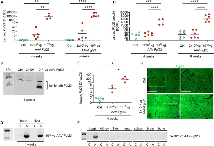
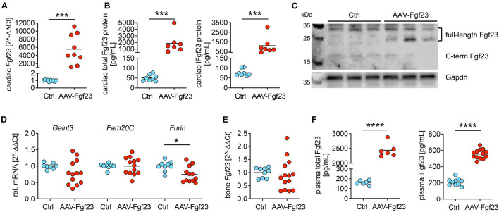
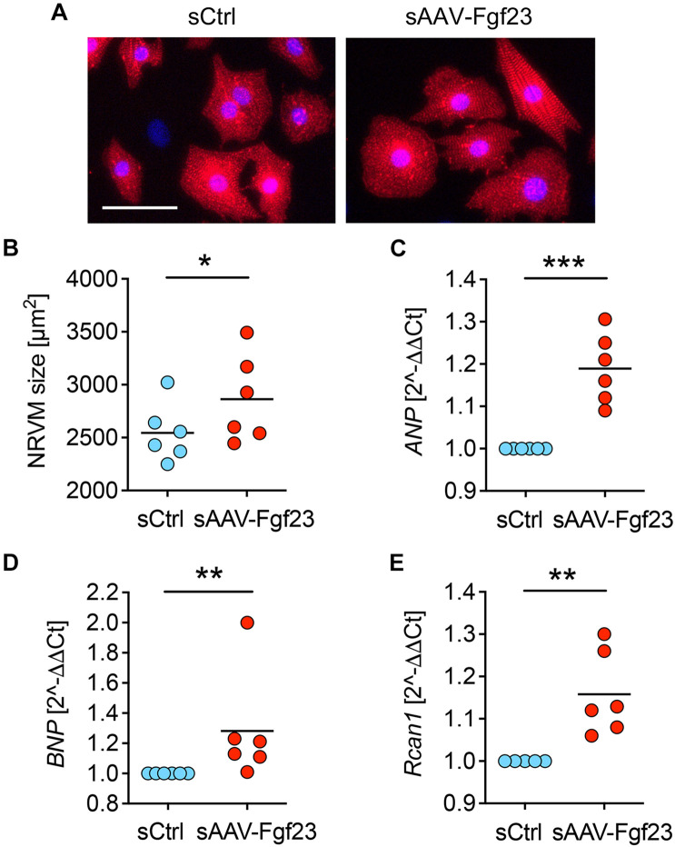
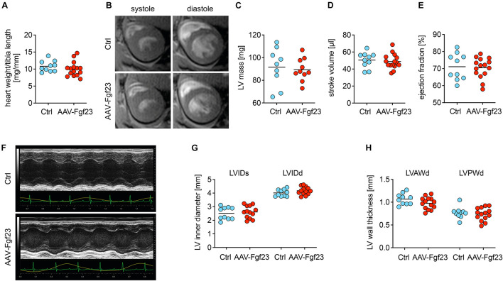
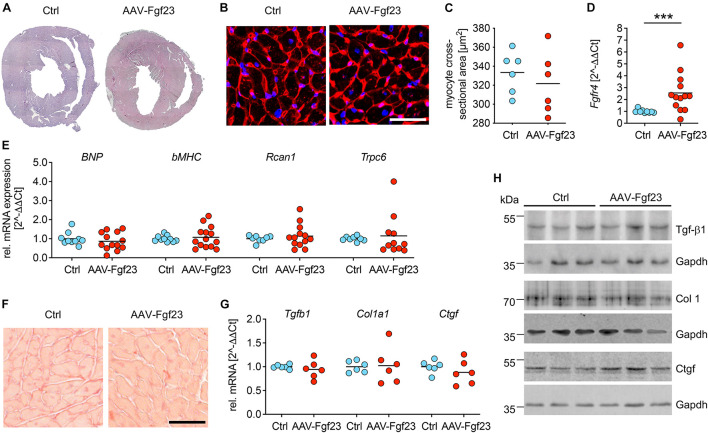
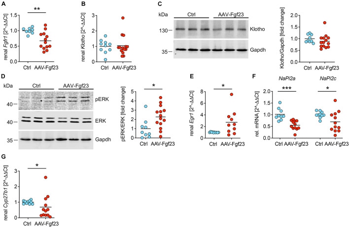
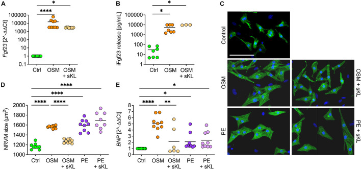
Fibroblast growth factor (FGF) 23 is elevated in chronic kidney disease (CKD) to maintain phosphate homeostasis. FGF23 is associated with left ventricular hypertrophy (LVH) in CKD and induces LVH via klotho-independent FGFR4-mediated activation of calcineurin/nuclear factor of activated T cells (NFAT) signaling in animal models, displaying systemic alterations possibly contributing to heart injury. Whether elevated FGF23 per se causes LVH in healthy animals is unknown. By generating a mouse model with high intra-cardiac Fgf23 synthesis using an adeno-associated virus (AAV) expressing murine Fgf23 (AAV-Fgf23) under the control of the cardiac troponin T promoter, we investigated how cardiac Fgf23 affects cardiac remodeling and function in C57BL/6 wild-type mice. We report that AAV-Fgf23 mice showed increased cardiac-specific Fgf23 mRNA expression and synthesis of full-length intact Fgf23 (iFgf23) protein. Circulating total and iFgf23 levels were significantly elevated in AAV-Fgf23 mice compared to controls with no difference in bone Fgf23 expression, suggesting a cardiac origin. Serum of AAV-Fgf23 mice stimulated hypertrophic growth of neonatal rat ventricular myocytes (NRVM) and induced pro-hypertrophic NFAT target genes in klotho-free culture conditions in vitro. Further analysis revealed that renal Fgfr1/klotho/extracellular signal-regulated kinases 1/2 signaling was activated in AAV-Fgf23 mice, resulting in downregulation of sodium-phosphate cotransporter NaPi2a and NaPi2c and suppression of Cyp27b1, further supporting the bioactivity of cardiac-derived iFgf23. Of interest, no LVH, LV fibrosis, or impaired cardiac function was observed in klotho sufficient AAV-Fgf23 mice. Verified in NRVM, we show that co-stimulation with soluble klotho prevented Fgf23-induced cellular hypertrophy, supporting the hypothesis that high cardiac Fgf23 does not act cardiotoxic in the presence of its physiological cofactor klotho. In conclusion, chronic exposure to elevated cardiac iFgf23 does not induce LVH in healthy mice, suggesting that Fgf23 excess per se does not tackle the heart.

Keywords: FGF23; fibrosis; klotho; left ventricular hypertrophy; mineral metabolism; mouse model.


Conflict of interest statement

TT filed and licensed patents in the field of non-coding RNAs and is the founder and shareholder of Cardior Pharmaceuticals GmbH. The remaining authors declare that the research was conducted in the absence of any commercial or financial relationships that could be construed as a potential conflict of interest.

Figures

FIGURE 1
FIGURE 1
Dose- and time-dependent evaluation of AAV-Fgf23. (A) Relative cardiac Fgf23 mRNA expression is dose-dependently increased in AAV-Fgf23 mice compared to Ctrl, irrespective of the duration of exposure. (B) ELISA-based quantification of cardiac Fgf23 protein in heart tissue lysates shows increased intact Fgf23 (iFgf23) protein only in AAV-Fgf23 mice administered with 1012 vector genomic particles (vg) compared to Ctrl. (C) Representative immunoblotting of Fgf23 in cardiac tissue demonstrates increased iFgf23 protein in the 1012 vg AAV-Fgf23 group after 4 weeks compared to Ctrl. (D) Semi-quantitative PCR targeting AAV vector shows excessive virus transduction after 4 weeks in the heart and the liver of 1012 vg AAV-Fgf23 mice (A) compared to Ctrl (C). (E) Hepatic Fgf23 mRNA expression is significantly increased after 4 weeks in 1012 vg AAV-Fgf23 mice compared to Ctrl. (F) By injecting 5 × 10¹¹ vg AAV-Fgf23, semi-quantitative PCR targeting AAV vector shows distinct virus transduction in the heart compared to Ctrl, whereas transduction in liver tissue was very low and absent in kidney, lung, spleen, brain, and bone tissue. (G) Immunofluorescent staining of cardiac mid-chamber sections demonstrates uniform Fgf23 (green) overexpression in almost all cardiac myocytes of the left ventricle in AAV-Fgf23 mice compared to Ctrl (original magnification ×20; scale bar, 500 and 250 μm, respectively). Data are given as scatter dot plots with means; *p < 0.05, **p < 0.01, ***p < 0.001, and ****p < 0.0001 analyzed using one-way ANOVA or Kruskal–Wallis test followed by Sidak’s or Dunn’s multiple comparisons post hoc tests, respectively, according to Shapiro–Wilk normality test; n = 3–7 mice per group.
FIGURE 2
FIGURE 2
Myocardial gene transfer of AAV-Fgf23 causes cardiac-specific Fgf23 overexpression resulting in the synthesis of intact Fgf23 protein. (A) Relative cardiac Fgf23 mRNA expression increases in AAV-Fgf23 mice compared to Ctrl. (B) ELISA-based quantification of cardiac Fgf23 protein in heart tissue lysates shows increased total and intact Fgf23 (iFgf23) protein in AAV-Fgf23 mice compared to Ctrl. (C) Representative immunoblotting of Fgf23 in cardiac tissue of three mice each demonstrates increased iFgf23 protein in AAV-Fgf23 mice compared to Ctrl, whereas C-terminal fragments are slightly reduced. Gapdh serves as the loading control. (D) Relative mRNA expression of Galnt3 and Fam20C is unchanged in AAV-Fgf23 mice compared to Ctrl, while Furin transcription is significantly reduced. (E) Fgf23 mRNA expression in the bone is similar between AAV-Fgf23 and Ctrl mice. (F) Plasma C-term and iFgf23 levels are significantly enhanced in AAV-Fgf23 mice compared to Ctrl. Data are given as scatter dot plots with means; *p < 0.05, ***p < 0.001, and ****p < 0.0001 analyzed using unpaired t-test or Mann–Whitney test according to Shapiro–Wilk normality test; n = 6–14 mice per group.
FIGURE 3
FIGURE 3
Serum of AAV-Fgf23 mice induces cardiac hypertrophy of NRVM. (A) Representative immunofluorescence images of isolated neonatal rat ventricular myocytes (NRVM) stimulated with 2% serum from AAV-Fgf23 (sAAV-Fgf23) or Ctrl (sCtrl) mice for 48 h. Myocytes are labeled with anti-α-actinin (red) and nuclei are counterstained with DAPI (blue) (original magnification ×20; scale bar 50 μm). (B) Quantification of the cross-sectional area of NRVM reveals hypertrophic cell growth due to treatment with sAAV-Fgf23 compared to sCtrl (mean of 100 cells per condition and isolation). (C–E) Quantitative real-time PCR analysis shows increased expression of ANP, BNP, and Rcan1 in sAAV-Fgf23-treated NRVM compared to sCtrl. Data are given as scatter dot plots with means; *p < 0.05, **p < 0.01, ***p < 0.001 analyzed using unpaired t-test or Mann–Whitney test according to Shapiro–Wilk normality test; n = 6 independent isolations of NRVM.
FIGURE 4
FIGURE 4
Chronic intra-cardiac Fgf23 synthesis does not impair cardiac function in unchallenged mice. (A) The heart weight to tibia length ratio is not increased in AAV-Fgf23 mice compared to Ctrl. (B) Representative cross-section MRI images of an AAV-Fgf23 and Ctrl mouse during systole and diastole. (C–E) Analyzed by MRI, left ventricular (LV) mass, stroke volume, and ejection fraction show no significant differences in AAV-Fgf23 mice compared to Ctrl. (F) Representative PLAX M-mode echocardiography images of an AAV-Fgf23 and Ctrl mouse. (G,H) In echocardiography, LV inner diameter during systole and diastole (LVIDs/d) and LV wall thicknesses during diastole (LV anterior wall, LVAW; LV posterior wall, LVPW) are not altered in AAV-Fgf23 mice compared to Ctrl. Data are given as scatter dot plots with means; n = 9–15 mice per group.
FIGURE 5
FIGURE 5
Adeno-associated virus expressing murine Fgf23 mice do not show any signs of pathological LVH. (A) Representative cross-sections of AAV-Fgf23 and Ctrl mice stained with HE (original magnification ×10). (B) Representative cross-sections of AAV-Fgf23 and Ctrl mice stained and wheat germ agglutinin (WGA) (original magnification ×40; scale bar, 50 μm). (C) Quantification of at least 100 individual cardiac myocytes per mouse reveals no size differences between both groups. (D) As analyzed by quantitative real-time PCR, cardiac Fgfr4 mRNA expression is significantly enhanced in AAV-Fgf23 mice compared to Ctrl. (E) The pro-hypertrophic NFAT target genes BNP, bMHC, Rcan1, and Trpc6 are not induced in AAV-Fgf23 mice compared to Ctrl. (F) Representative images of picrosirius red-stained mid-chamber free-wall of AAV-Fgf23 and Ctrl mice (original magnification, ×63; scale bar, 50 μm). (G,H) The mRNA and protein expression of fibrosis-associated markers Tgfb1, Col1a1, and Ctgf and are not altered in AAV-Fgf23 mice. Gapdh serves as the loading control. Data are given as scatter dot plots with means; ***p < 0.001 analyzed using Mann–Whitney test according to D’Agostino and Pearson’s normality test; n = 6–14 mice per group.
FIGURE 6
FIGURE 6
Enhanced secretion of cardiac iFgf23 activates FGFR1/klotho/ERK signaling in the kidney. (A) Analyzed by quantitative real-time PCR, renal expression of Fgfr1 is decreased in AAV-Fgf23 mice compared to Ctrl. (B) Renal Klotho mRNA levels are equal in both groups. (C) Representative immunoblots followed by quantification verify normal klotho protein levels in kidney tissue of AAV-Fgf23 mice compared to Ctrl. Gapdh serves as the loading control. (D) Representative immunoblots in kidney tissue followed by quantification show increased phosphorylation of ERK1/2 in AAV-Fgf23 mice compared to Ctrl with no changes of total ERK1/2 protein. Gapdh serves as the loading control. (E) Renal mRNA expression of Egr1 increases in AAV-Fgf23 mice compared to Ctrl, confirming AAV-Fgf23 mediated activation of ERK signaling pathway. (F) Renal mRNA expression of NaPi2a and NaPi2c decreases in AAV-Fgf23 mice compared to Ctrl. (G) mRNA expression of Cyp27b1 is downregulated in AAV-Fgf23 mice compared to Ctrl. Data are given as scatter dot plots with means; *p < 0.05, **p < 0.01, and ***p < 0.001 analyzed using unpaired t-test or Mann–Whitney test according to D’Agostino and Pearson’s normality test; n = 8–14 mice per group.
FIGURE 7
FIGURE 7
Soluble klotho suppresses cardiac FGF23-induced cardiac hypertrophy in vitro. (A) Stimulation with oncostatin M (OSM) increases endogenous Fgf23 mRNA expression in isolated NRVM irrespective of cotreatment with soluble klotho (sKL). (B) ELISA-based quantification of intact Fgf23 (iFgf23) in conditioned medium of NRVM reveals enhanced iFgf23 release due to OSM treatment, while sKL costimulation does not alter OSM-mediated high iFgf23 secretion. (C,D) Immunofluorescence images of isolated NRVM and quantification of cross-sectional cell area observe hypertrophic growth of OSM stimulated NRVM. Phenylephrine (PE) serves as the positive control for cardiac hypertrophy. Cotreatment with sKL protects from cardiac hypertrophy induced by OSM but not by PE. Myocytes are labeled with anti-α-actinin (green) and nuclei are counterstained with DAPI (blue) (original magnification ×20; scale bar 100 μm). (E) Quantitative real-time PCR analysis shows increased expression of BNP in OSM and PE-treated NRVM compared to Ctrl that is only blocked by sKL in the OSM group. Data are given as scatter dot plots with means; p < 0.05, ∗∗p < 0.01, and *⁣*⁣**p < 0.0001 analyzed using one-way ANOVA or Kruskal–Wallis test followed by Sidak’s or Dunn’s multiple comparisons post hoc tests, respectively, according to Shapiro–Wilk normality test; n = 6–9 independent isolations of NRVM; conditioned medium was used from n = 3–7 independent NRVM isolations.

References

    1. Andrukhova O., Slavic S., Odörfer K. I., Erben R. G. (2015). Experimental myocardial infarction upregulates circulating fibroblast growth factor-23. J. Bone Miner. Res. 30 1831–1839. 10.1002/jbmr.2527 - DOI - PMC - PubMed
    1. Andrukhova O., Slavic S., Smorodchenko A., Zeitz U., Shalhoub V., Lanske B., et al. (2014). FGF23 regulates renal sodium handling and blood pressure. EMBO Mol. Med. 6 744–759. - PMC - PubMed
    1. Bao J. F., Hu P. P., She Q. Y., Li A. (2020). A land of controversy: fibroblast growth factor-23 and uremic cardiac hypertrophy. J. Am. Soc. Nephrol. 31 1423–1434. 10.1681/asn.2020010081 - DOI - PMC - PubMed
    1. Barth J. H., Jones R. G., Payne R. B. (2000). Calculation of renal tubular reabsorption of phosphate: the algorithm performs better than the nomogram. Ann. Clin. Biochem. 37 79–81. 10.1258/0004563001901371 - DOI - PubMed
    1. Böckmann I., Lischka J., Richter B., Deppe J., Rahn A., Fischer D. C., et al. (2019). FGF23-mediated activation of local RAAS promotes cardiac hypertrophy and fibrosis. Int. J. Mol. Sci. 20:4634. 10.3390/ijms20184634 - DOI - PMC - PubMed